Rhorus Förster, 1869

Taxonomic History / Nomenclature
Rhorus Foerster, 1869: 195. Type species: Tryphon mesoxanthus Gravenhorst, 1829. Included by Woldstedt (1877: 455). Monobasic.
Dolichoblastus Strobl, 1903: 52. Type species: Monoblastus (Dolichoblastus) flavopictus Strobl. Monobasic. Synonymized by Townes (1945: 485).
Cyphanza Cameron, 1909: 723. Type species: Cyphanzia nigra Cameron. (nigra preoccupied in Rhorus by Ashmead, 1902; = cameroni Townes, 1970). Monobasic.

Cyphanza was treated by Townes (1945) as a subgenus of Rhorus, containing virtually all of the smaller Nearctic species. Townes (1945) reserved Rhorus s.s. for the large-bodied Nearctic species clapini, which closely resembles the type species of Rhorus, R. mesoxanthus. Barron (1986) delineated six species groups in his revision of the Nearctic species and did not recognize subgenera.

Remarks
50 valid species were listed by Yu et al. (2012). Gauld (1997) stated that this was the most speciose genus of Pionini. The Nearctic species were revised by Barron (1986).
Diagnosis and Relationships
Rhorus is usually and perhaps best recognized by the absence of an epistomal sulcus separating the face from the clypeus (Figs 1, 2), though in males of some species, there is a weak but distinct impression. The ovipositor is short, straight, and directed dorsally at rest (Fig. 3). Rhorus is further characterized by the well-developed glymma, pectinate claws, and by having the dorsal end of epicnemial carina always distinctly ending at the anterior margin of the mesopleuron.

Rhorus clapini (Provancher) from the Nearctic is very similar to Rhorus mesoxanthus (Grav.) from Europe. Both are very large and have exactly the same yellow ovipositor sheath. They seem to differ primarily in the development of the T1 carinae. These large-bodied species of Rhorus can be confused with Ctenopelma because of the there is a carina extending from the base of T2 laterally to the T2 spiracle, but the glymma is smaller in Rhorus and clypeus is different.

324_mximage
1. Rhorus face and clypeus...
322_mximage
2. Rhorus face and clypeus...
326_mximage
3. Rhorus ovipositor...
 
Description
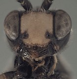
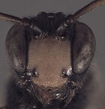
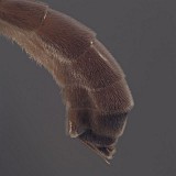
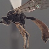
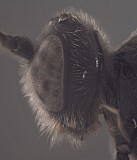
Clypeus (Fig. 3) with surface more or less uniformly setose and punctate to granular-punctate; ventral margin varying from evenly and distinctly convex to weakly convex and nearly truncate to nearly dentate or angled forward medially and appearing slightly emarginate when the head is tilted backwards as in Fig. 4; margin blunt; epistomal sulcus usually absent, broadly and shallowly impressed, especially in males, in a few species; clypeus in profile flat to weakly protruding. Face flat to weakly bulging in profile; setose, densely punctate to granular-punctate; inner eye margins more or less parallel. Ocelli small, diameter of lateral ocellus much less than distance from ocellus to eye. Malar space distinct, varying from a little less than half basal width of mandible to nearly equal basal width of mandible; malar sulcus absent. Mandible (Fig. 4) broad, gradually narrowing from base to apex; weakly to strongly curved; dorsal tooth usually equal to or slightly larger than ventral tooth, rarely with ventral tooth larger; basal, transverse impression well-developed; ventral margin distinctly carinate. Maxillary palp a little shorter than height of head; female antenna equal to or slightly shorter than body; equal in length to or longer than body in male. Hypostomal carina meeting occipital carina distinctly above base of mandible; occipital carina complete. Epomia well-developed in some species, obscured by surrounding sculpture in others and either absent or difficult to discern. Epicnemial carina reaching anterior margin of mesopleuron. Notaulus variable: commonly absent, broad and shallow in some species, sometimes marked by more distinct punctation extending posteriorly past level of tegula and converging to a very broad, very shallow, median depression, distinctly impressed anteriorly in a few species. Groove between propodeum and metapleuron broadly u-shaped, readily visible in lateral view in most species, less so in a few species, where it appears more v-shaped; broad, u-shaped groove mid-dorsally between propodeum and metanotum present and usually readily visible in lateral view; pleural carina well-developed, complete; propodeal carinae variably developed, completely carinate, with well-delineated areola, in some species; median longitudinal carinae always well developed, lateral longitudinal carina sometimes poorly developed to absent anteriorly, posterior transverse carina usually complete but sometimes absent or indistinct laterally, anterior transverse carina often absent laterally and/or medially, with areola and median basal area confluent; petiolar area usually with median longitudinal carina. Apical margin of mid tibia expanded into a distinct tooth similar to that of fore leg; apical comb on posterior side of hind tibia absent; posterior hind tibial spur long, 0.3-0.5 times length of hind basitarsus; tarsal claws variously pectinate, densely and completely so on all legs in type species and the closely related 
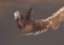
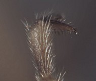
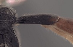
R. clapini, more sparsely so and often basally situated in many other species, as in Figs. 6 and 7. Fore wing areolet present, though often with large bulla on one side; stigma varying from relatively narrow as in the type species, with Rs+2r arising near basal 0.3 of stigma to broader and wedge or triangular in shape, with Rs+2r arising near middle or less commonly near basal 0.3-0.4. Hind wing with first abscissa of CU1 longer than 1cu-a, often very much longer. T1 (Fig. 8) longer and more slender in the type species and a few others, but more commonly shorter and relatively stout, gradually expanding from base to apex; straight in profile; dorsal carinae usually sharp and well developed at least to level of spiracle and often more posteriorly but rarely extending full length of T1; basal depression at dorsal tendon attachment deep, broad; dorsal-lateral carina usually complete between spiracle and apex of T1; glymma large, deep, basally situated. S1 extending to level of spiracle or nearly so in the type species and a few others, extending only about 0.3 times length of T1 in many species. T2 thyridium absent, at least one species with a well-developed carina between anterior margin of T2 and spiracle; laterotergites of T2 and T3 separated by creases from median tergite. Ovipositor (Figs 9, 10) straight; ovipositor and sheath usually directed dorsally at rest; ovipositor relatively shorter than in Pion, somewhat gradually becoming needle-like apically from distinctly swollen base (Fig. 10); without dorsal, subapical notch.

The above description is greatly modified from Townes (1970) and is based on several species in the Texas A&M University collection. A detailed description is also presented by Gauld (1997).

320_mximage
1.Rhorus
325_mximage
2.Rhorus habitus
322_mximage
3.Rhorus face
324_mximage
4. Rhorus face and mandibl...
327_mximage
5. Rhorus head lateral...
343_mximage
6.Rhorus tarsal claws
346_mximage
7.Rhorus claws
328_mximage
8.Rhorus T1 lateral
326_mximage
9. Rhorus apex of metasoma,...
6684_mximage
10. Rhorus reproductive trac...
 
Distribution
Mostly known from the Holarctic, but Gauld (1997) described one species from southern Costa Rica and also noted that it occurs in the Oriental region where it borders the Palaearctic.
Distribution
No referenced distribution records have been added to the database for this OTU.
Biology / Hosts
A parasitoid of sawflies, ovipositing through the larval stemmata Pschorn-Walcher and Zinnert (1971). Eller et al. (1989) recorded two species as parasitoids of Pikonema alaskensis (Rohwer) in their Minnesota (USA) sample site, and discussed diapause in the host and its parasitoids. Eichhorn and Pschorn-Walcher (1978) provided information on levels of parasitism and general ecology for European populations of Pristiphora geniculata.
Map

There are no specimens currently determined for this OTU, or those specimens determined for this OTU are not yet mappable.

Acknowledgements
This page was assembled by Bob Wharton as part of a larger collaborative effort on the genera of Ctenopelmatinae.

This work would not have been possible without the groundwork provided by Ian Gauld’s study of the Australian and Costa Rican faunas, and we are particularly grateful for his assistance in many aspects of this study. We also thank David Wahl of the American Entomological Institute and Andy Bennett of the Canadian National Collection for extended loans of the material used for this study and particularly Dave Karlsson for sending valuable material from the Swedish Malaise Trap Survey (trap 20, collection event 233) and Al Gillogly for material from Idaho. Matt Yoder provided considerable assistance with databasing issues, and our use of PURLs (http://purl.oclc.org) in this regard follows the example of their use in publications by Norm Johnson. Heather Cummins, Andrea Walker, Patricia Mullins, Caitlin Nessner, Amanda Ladigo, Jacques Dubois and Cheryl Hyde graciously assisted with image processing, formatting, and literature retrieval. This study was supported by the National Science Foundation’s PEET program under Grant No. DEB 0328922 and associated REU supplement #s DEB 0723663, 0923134, and 1026618. Page last updated April, 2015.

This material is based upon work at Texas A&M University supported by the National Science Foundation under Grant Number DEB 0328922 with REU supplements DEB 0723663, 0923134, and 1026618. Any opinions, findings, and conclusions or recommendations expressed in this material are those of the author(s) and do not necessarily reflect the views of the National Science Foundation.