Syntactus Förster, 1869

Taxonomic History / Nomenclature
Syntactus Foerster, 1869: 210.
Type species: Ichneumon delusor Linnaeus, 1758. Subsequently designated by Perkins (1962: 456).

Tromopoea Foerster, 1869: 210.
Type species: Catoglyptus minor Holmgren, 1857. Subsequently designated by Perkins (1962: 460). Synonymized by Perkins (1962: 460)

Brischkea Kriechbaumer, 1897: 167.
Type species: Brischkea parvula Kriechbaumer, 1897 (a junior synonym of S. delusor (Linnaeus). Monobasic. Genus and species synonymized by Perkins (1962: 456).

Remarks
The following valid species were included by Yu et al. (2012).

Syntactus delusor (Linnaeus, 1758)
Syntactus fusiformis (Thomson, 1894)
Syntactus julianicus Sun and Sheng, 2012
Syntactus leleji Kasparyan, 2007
Syntactus minor (Holmgren, 1857)
Syntactus minutus (Bridgman, 1886)
Syntactus varius (Holmgren, 1858)

Diagnosis and Relationships
The first metasomal tergite in Syntactus is similar in shape to that found in the species of Pion and both lack a glymma on the distinctive T1 in which the narrow and parallel-sided basal portion extends past the spiracles. The first metasomal sternite is also long in both: extending posteriorly to the spiracles. The mandibles, with the distinctly carinate ventral margin, are relatively shorter than in Pion and the clypeus is not impressed along its apical margin in Syntactus. See also comments in the diagnosis section for Rhaestus.
Description
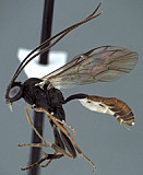
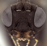
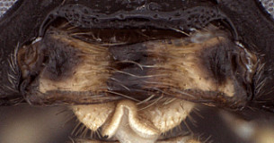
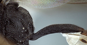
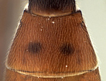
Clypeus with surface more or less uniformly setose and finely rugulose (Fig. 2); ventral margin evenly but weakly convex, the margin blunt but not broadly so (Fig. 2); epistomal sulcus impressed throughout, weaker medially; clypeus in profile weakly protruding (Fig. 1). Face setose, densely granular-punctate medially, faintly punctate, nearly smooth laterally as well as on frons and vertex; inner eye margins weakly (Fig. 2) to distinctly converging. Malar space short but distinct, about half basal width of mandible in female, slightly longer in male; malar sulcus absent. Mandible (Fig. 3) broad, nearly parallel-sided; dorsal tooth distinctly narrower and a little shorter than ventral tooth; basal, transverse impression absent; ventral margin distinctly carinate. Maxillary palp a little shorter than height of head; female antenna slightly shorter than body; about equal in length to body in male. Hypostomal carina meeting occipital carina at base of mandible or the two very slightly separated at the base; occipital carina complete. Epicnemial carina reaching anterior margin of mesopleuron. Notaulus distinct, varying from weakly to deeply impressed, sculptured when deeply impressed; extending posteriorly to level of tegula. Groove between propodeum and metapleuron broadly u-shaped, readily visible in lateral view; broad, u-shaped groove mid-dorsally between propodeum and metanotum also readily visible in lateral view; pleural carina well-developed, complete; median and lateral longitudinal carinae well developed anteriorly, variously effaced posteriorly, though rarely completely so; posterior transverse carina sometimes complete laterally, usually absent medially, anterior transverse carina present medially absent laterally, thus areola usually open posteriorly. Hind femur stout; apical margin of mid tibia not expanded into a distinct tooth similar to that of fore leg; apical comb on posterior side of hind tibia absent; posterior hind tibial spur about 7x longer than maximum width at base; tarsal claws smaller than in 
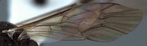
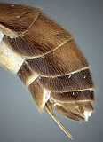
Pion, not pectinate. Fore wing areolet absent; stigma relatively narrow (Fig. 1), Rs+2r arising near basal 0.3-0.4 of stigma. Hind wing (Fig. 4) with first abscissa of CU1 varying from equal in length to slightly shorter or longer than 1cu-a. T1 (Fig. 5) long, slender, parallel-sided from base to distinctly posteriorad spiracles, then abruptly expanding; weakly decurved in profile (Fig. 5); dorsal carinae extending full length of parallel-sided portion (to posterior end of S1), fading into and largely absent from expanded posterior portion; basal depression at dorsal tendon attachment small; dorsal-lateral carina complete between spiracle and apex of T1; glymma absent. S1 extending full length of parallel-sided portion (over basal 0.6, distinctly posteriorad spiracle). T2 thyridium absent (Fig 6); laterotergites of T2 and T3 separated by creases from median tergite. Ovipositor (Fig. 7) straight; ovipositor sheath more distinctly upcurved; ovipositor relatively shorter than in Pion, abruptly becoming needle-like apically from distinctly swollen base (Fig. 7); without dorsal, subapical notch.

This description is based largely on S. minor and is expanded from that given for Syntactus by Townes (1970), who also included it in a key to genera of Pionini.

10011_mximage
1.
10007_mximage
2. Syntactus minor face...
10008_mximage
3. Syntactus minor mandible...
10009_mximage
4.Syntactus minor wing
10005_mximage
5.Syntactus minor T1
10006_mximage
6.Syntactus minor T2
10004_mximage
7. Syntactus minor...
 
Distribution
Eastern and Western Palaearctic, with one species described from Jiangxi, China. Townes (1970) noted two undescribed species from Japan and China.
Distribution
No referenced distribution records have been added to the database for this OTU.
Biology / Hosts
The following hosts and references for those hosts records are from Yu et al. (2005).

Lithosia quadra (Hedwig 1944, 1950)
Rhyacionia resinella (Hedwig 1951; Fulmek 1968)
Strongylogaster macula (Bauer 1958)

The record from Rhyacionia, a lepidopteran, is suspect and needs verification.

Map

There are no specimens currently determined for this OTU, or those specimens determined for this OTU are not yet mappable.

Acknowledgements
This page was assembled by Bob Wharton as part of a larger collaborative effort on the genera of Ctenopelmatinae.

This work would not have been possible without the groundwork provided by Ian Gauld’s study of the Australian and Costa Rican faunas, and we are particularly grateful for his assistance in many aspects of this study. We also thank David Wahl of the American Entomological Institute and Andy Bennett of the Canadian National Collection for extended loans of the material used for this study and particularly Dave Karlsson for sending valuable material from the Swedish Malaise Trap Survey (trap 37, collection event 831). Matt Yoder provided considerable assistance with databasing issues, and our use of PURLs (http://purl.oclc.org) in this regard follows the example of their use in publications by Norm Johnson. Heather Cummins, Andrea Walker, Patricia Turner, Caitlin Nessner, Amanda Ladigo, and Cheryl Hyde graciously assisted with image processing, formatting, and literature retrieval. This study was supported by the National Science Foundation’s PEET program under Grant No. DEB 0328922 and associated REU supplement #s DEB 0723663, 0923134, and 1026618. Page last updated Jan, 2015.

This material is based upon work supported by the National Science Foundation under Grant Number DEB 0328922 with REU supplements DEB 0723663, 0923134, and 1026618. Any opinions, findings, and conclusions or recommendations expressed in this material are those of the author(s) and do not necessarily reflect the views of the National Science Foundation.