Lathiponus Foerster, 1868

Taxonomic History / Nomenclature
Lathiponus Foerster, 1869: 198. Type species: Mesoleius (Lathiponus) pulcherrimus Thomson, 1888 [a junior subjective synonym of Bassus frigidus Woldstedt, 1874, species synonymized by Hellen 1953]. Subsequent inclusion by Thomson (1888): 1261. (Monobasic.)

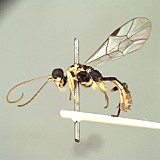
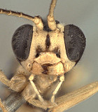
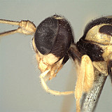
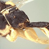
Polyselasmus Schmiedeknecht, 1912: 2552. Type species: Eclytus semiluctuosus Vollenhoven, 1878 [a junior subjective synonym of Bassus frigidus Woldstedt, 1874]. Monobasic.

Ceratosaotis Gregor, 1939: 85. Type species: Ceratosaotis ornatus Gregor, 1939 [a junior subjective synonym of Bassus frigidus Woldstedt, 1874, species synonymized by Townes 1970]. Original designation. Genus synonymized by Townes (1970).

Remarks
There is a single species in the genus, sometimes (e. g. Yu and Horstmann 1997) referred to as Lathiponus bicolor (Brischke, 1878), for which there are four junior, subjective synonyms, one of which (frigidus Woldstedt) is also a junior, primary homonym. Yu et al. (2005) suggest that the correct name for this species is Lathiponus semiluctuosus (Vollenhoven, 1878).

The above description is based on a single female specimen from Germany borrowed from The Natural History Museum, London, but closely matches the description provided by Townes (1970).

Diagnosis and Relationships
Lathiponus is one of several genera that were grouped by Gauld (1997) on the form of the glymma: the two sides extending medially towards the median, basal depression at the dorsal tendon attachment rather than meeting medially posterior to the dorsal tendon attachment. Lathiponus is defined by the deep impression posteriorly on the dorsal part of vertex and occiput that obliterates the dorsal-median part of the occipital carina, and by a bilobed, tuberculate process at the ventral part of this depression. This feature is not found in any other perilissine. The ovipositor and sheath, as figured above and by Townes (1970), are strongly upcurved. The overall shape of the clypeus, largely the result of the uneven ventral margin, is also distinctive among the perilissines.
Description
Clypeus with ventral margin sharp, weakly concave over median 0.3 then sharply angled dorsal-laterally on either side resulting in a vaguely trapezoidal outline (Fig. 3), somewhat toothed ventral-laterally, at least in female, on either side of medially concave part of margin; epistomal sulcus distinct, deep throughout. Malar space distinct, shorter than basal width of mandible (Fig. 4). Mandible with dorsal tooth about as long as ventral tooth (Fig. 3). Ocelli small, lateral ocellus distinctly shorter than distance between ocellus and eye. Maxillary palp about equal in length to height of head; female antennae about as long as body or slightly longer (Figs 1, 2); first flagellomere with discrete tyloid not apparent. Hypostomal carina not joining occipital carina above base of mandible; occipital carina absent medially where occiput extends narrowly forward to ocellar triangle (Fig. 5). Dorsal end of epicnemial carina distant from anterior margin of mesopleuron. Notaulus absent. Neither small v-shaped notch nor distinct u-shaped notch present between propodeum and metanotum in lateral view. Pleural carina complete, well-developed; propodeal carinae well-developed medially, weaker laterally, with part of posterior and nearly all of anterior transverse carinae absent laterally or reduced to indistinct wrinkled sculpture (Fig. 6), areola very narrow anteriorly, somewhat trapezoidal to triangular, median posterior area sharply delimited. Apical margin of mid tibia expanded into a distinct tooth similar to that on fore tibia; posterior hind tibial spur at least 7x longer than maximum width at base; apical comb of setae on posterior face of hind tibia not well developed; tarsal claws pectinate though not strongly so, most evident on fore leg. Fore wing areolet absent; Rs+2r arising slightly basad midpoint of stigma. Hind wing with first abscissa of CU1 much longer than 1cu-a, the latter exceptionally short and distal abscissa of CU1 barely indicated. T1 not long and slender, distinctly broadening posteriorly, dorsal carinae weak, indistinct anteriorly, absent posteriorly; basal depression at dorsal tendon attachment deep, distinct; glymma (Fig. 6) shallow but very large, extending medially towards median basal depression and posteriorly to spiracle. T2 thyridium absent; laterotergites of T2 and T3 not separated by sharp creases. Ovipositor with deep, broad subapical notch (Fig. 7); ovipositor sheath relatively short, strongly upcurved as in Rhorus. No males seen.
18415_mximage
1. Lathiponus lateral habit...
18416_mximage
2. Lathiponus lateral habit...
18414_mximage
3. Lathiponus face show...
18420_mximage
4. Lathiponus head in profi...
18441_mximage
5. Lathiponus back of head showing do...
18418_mximage
6. Lathiponus propodeal and...
18417_mximage
7. Lathiponus showing ov...
 
Distribution
The only known species occurs in Europe.
Distribution
No referenced distribution records have been added to the database for this OTU.
Biology / Hosts
The type species has been recorded from several species of Tenthredinidae.
Map

There are no specimens currently determined for this OTU, or those specimens determined for this OTU are not yet mappable.

Acknowledgements
This page was assembled by Bob Wharton as part of a larger collaborative effort on the genera of Ctenopelmatinae. This work would not have been possible without the groundwork provided by Ian Gauld’s study of the Australian and Costa Rican faunas, and we are particularly grateful for his assistance in many aspects of this study. We also thank David Wahl for useful feedback throughout our study and Gavin Broad for the loan of the specimen used in the images and description, as well as exchange of information on Perilissini. Matt Yoder provided considerable assistance with databasing issues, and our use of PURLs (http://purl.oclc.org) in this regard follows the example of their use in publications by Norm Johnson. Caitlin Nessner and Amanda Ladigo graciously assisted with formatting and literature retrieval and Lauren Ward did the imaging. This study was supported by the National Science Foundation’s PEET program under Grant No. DEB 0328922 and associated REU supplement nos DEB 0723663 and 1026618. Page last updated September, 2011.

This material is based upon work supported by the National Science Foundation under Grant Number DEB 0328922 with REU supplements DEB 0723663 and 1026618.
Any opinions, findings, and conclusions or recommendations expressed in this material are those of the author(s) and do not necessarily reflect the views of the National Science Foundation.