Trematopygodes Aubert, 1968
Taxonomic History / Nomenclature
Trematopygodes Aubert, 1968: 69. Type species: Trematopygus aprilinus Giraud, 1872. Monobasic.
Remarks
Seventeen valid species were included by Yu et al. (2005). Trematopygodes was recently revised for the Holarctic region by Hinz and Horstmann (1998), an expansion of the earlier work on European species by Hinz (1980).
Diagnosis and Relationships

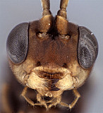
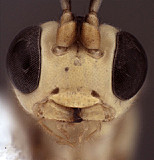
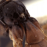
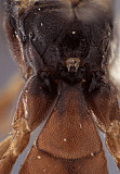
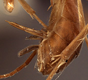
Defining features include the lateral, triangular to rounded lobes along the ventral margin of the clypeus (present in females: Fig. 1 but absent in males: Fig. 2), posteriorly broadening first metasomal tergite (Fig. 3) with typical perilissine glymma (Fig. 4) and the dorsal tendon attachment set within a weak but distinct basal depression. The metasomal terga are usually sculptured throughout in females. Lateral lobes are present along the ventral margin of the clypeus in other genera, such as Oetophorus and Aechmeta, but are particularly well-developed in most species of Trematopygodes.
Description
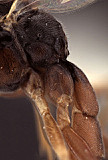
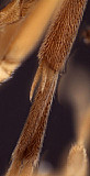
Clypeus with ventral margin thick and bluntly rounded, with thin, usually prominent, triangular to rounded tooth laterally in female (Fig. 5), this absent in male (Fig. 6); ventral margin convex; epistomal sulcus distinct throughout, clypeus in profile distinctly angled outwardly from face. Malar space distinct, usually at least half basal width of mandible. Mandible with ventral tooth usually very slightly longer than dorsal tooth, more rarely with dorsal tooth distinctly longer than ventral tooth. Ocelli small, lateral ocellus distinctly shorter than distance between ocellus and eye. Maxillary palp shorter than head height; female antennae varying from about as long as to distinctly shorter than body; first flagellomere with discrete tyloid containing fewer than 15 sensilla. Hypostomal carina joining occipital carina well above base of mandible; occipital carina complete. Dorsal end of epicnemial carina distant from anterior margin of mesopleuron. Notaulus absent to barely indicated as a broad, very shallow depression at anterior margin, not extending posteriorly to level of spiracle. U- to v-shaped groove or notch absent between propodeum and metanotum in lateral view; pleural carina complete, well-developed; propodeal carinae (Figs 8-10) well-developed posteriorly with complete posterior transverse carina and petiolar and posterior-lateral fields, absent or nearly so anteriorly, areola at most indistinct, anterior transverse carina absent, lateral longitudinal carina sometimes weakly indicated. Apical margin of mid tibia weakly to sharply expanded into a distinct tooth similar to that on fore tibia; apical comb on hind tibia not well developed, row of setae present along posterior face, but these widely spaced, not dense as in typical comb; posterior hind tibial spur not exceptionally long (Fig. 11), but at least 7x longer than maximum width at base; tarsal claws completely pectinate. Fore wing (Fig. 7) with areolet present, usually broad; stigma relatively broad, Rs+2r arising at or near midpoint. Hind wing with first abscissa of CU1 distinctly longer than 1cu-a. T1 (Figs 1, 3, 8-10) not long and slender throughout, distinctly broadening posteriorly, usually without dorsal carinae, but with weakly elevated, rounded ridges in at least one species; usually with shallow but distinct basal depression at dorsal tendon attachment; dorsal-lateral carina extending from spiracle to apex of T1 in some species, weak to absent in others; glymmae on each side meeting on the midline posterior to dorsal tendon attachment, deep, usually tall, separated at midline by translucent partition. T2 thyridium absent; laterotergites of T2 and T3 completely separated by creases. Ovipositor (Figs. 12-14) straight, relatively broad at base, with shallow, very broad subapical notch, the lower valves much deeper than upper valves over apical 0.6; ovipositor sheath long, narrow, slender, somewhat rounded distally. Male parameres broad, rectangular in dorsal view, never narrowly attenuate distally; aedeagus rounded and clubbed distally.
The description given above is based on four of the Nearctic species.
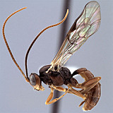
1.
Trematopygodes lateral h...
↴
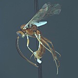
2.
Trematopygodes lateral h...
↴
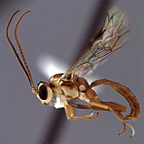
3.
Trematopygodes lateral h...
↴
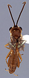
4.
Trema...
↴
5.
Trematopygodes face, ...
↴
6.
Trematopygodes face, m...
↴
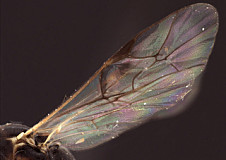
7.Trematopygodes wings.
8.
Trematopygodes...
↴
9.
Trematopygodes propodeum...
↴
10.
Trematopygodes ...
↴
11.
Trematopy...
↴
12.Trematopygodes ovipositor

13.
Trematopygodes oviposito...
↴
14.
Trematopygodes oviposito...
↴
Distribution
No referenced distribution records have been added to the database for this OTU.
Biology / Hosts
Host records are available for five of the described species of Trematopygodes. Four of these have been reared from hosts in the genus Periclista. One of the Nearctic species has also been reared from Eupareophora parva. All known hosts are blennocampine Tenthredinidae.
Map
There are no specimens currently determined for this OTU, or those specimens determined for this OTU are not yet mappable.
Acknowledgements
This page was assembled by Bob Wharton as part of a larger collaborative effort on the genera of Ctenopelmatinae. This work would not have been possible without the groundwork provided by Ian Gauld’s study of the Australian and Costa Rican faunas, and we are particularly grateful for his assistance in many aspects of this study. We also thank David Wahl for useful feedback throughout our study and to Gavin Broad for exchange of information on Perilissini. Matt Yoder provided considerable assistance with databasing issues, and our use of PURLs (http://purl.oclc.org) in this regard follows the example of their use in publications by Norm Johnson. Patricia Mullins, Andrea Walker, Amanda Ladigo, Heather Cummins, and Cheryl Hyde graciously assisted with image capture, processing, and formatting of both figures and text. This study was supported by the National Science Foundation’s PEET program under Grant No. DEB 0328922 and associated REU supplement nos DEB 0723663 and 1026618. Page last updated February, 2011.
This material is based upon work at Texas A&M University supported by the National Science Foundation under Grant Number DEB 0328922 with REU supplements DEB 0723663 and number 1026618. Any opinions, findings, and conclusions or recommendations expressed in this material are those of the author(s) and do not necessarily reflect the views of the National Science Foundation.
