Euryproctus Holmgren, 1857

Taxonomic History / Nomenclature
Euryproctus Holmgren (1857: 109). Type species: Mesoleptus annulatus Gravenhorst, 1829. Subsequent designation by Viereck (1912).

Sychnoleter Foerster, 1869: 197 Type species: Mesoleptus geniculosus Gravenhorst, 1829. Subsequently included and specifically designated by Thomson (1889: 1430). Monobasic. Synonymized by Townes et al. (1965: 269).

Hypocryptus Foerster, 1869: 198 Type species: Mesoleptus (Hypocryptus) testaceicornis Brischke, 1892 [= a junior subjective synonym of Euryproctus mundus Gravenhorst, 1829]. Subsequent inclusion by Brischke, 1892. Monobasic. Synonymized by Townes et al. (1965: 269).

Xenonastes Foerster, 1869: 208 Type species: Euryproctus alpinus Holmgren, 1857. Subsequent designation by Townes et al. (1965). Synonymized by Townes et al. (1965: 269).

Fovaya Cameron, 1903: 341. Type species: Fovaya annulicornis Cameron. Monobasic. Apparently first synonymized by Morley (1913).

The publication by Holmgren is often cited with different dates, most commonly 1855 in older literature.

Remarks
41 valid species were included by Yu et al. (2012). Euryproctus is largely Holarctic, with a few Oriental species known.

At least some of the species of Euryproctus exhibit extreme sexual dimorphism in color pattern as exemplified by the species in Figs. 2-4 (females in Figs 2 and 3, male in Fig. 4). In many others, the color differences are primarily limited to face and clypeus (usually dark in females and pale in males).

The clypeus, when examined across species, shows and interesting morphocline from an apical margin that is sharp throughout to one where there is a subapical bulge that barely overlaps the sharp margin medially, with the medial part of the sharp margin now appearing impressed below the bulge. In various species, the subapical bulge becomes larger and forms varying amounts of the middle part of the apical margin, this margin now blunt as the sharp margin is restricted laterally. At the other end of the morphocline, the apical margin is completely rounded (blunt). Without additional studies, the direction of the cline cannot be readily ascertained.

Diagnosis and Relationships
The species of Euryproctus are variable in some of the key characters used to separate groups of other genera in the Euryproctini, notably the fore wing areolet and the apical margin of the clypeus. The areolet is present in some species and absent in others. For most of the species examined, the areolet was generally (90-100% of wings) consistent in its presence or absence, but in a few species, it was more variable. The apical margin of the clypeus was usually blunt at least medially in most species examined, but sharp throughout in at least one species. Euryproctus is perhaps best distinguished by characteristics specific to females and males. Females are readily diagnosed by the characteristic tubular, posteriorly-directed to down-curved ovipositor sheath and the hypopygium with short, erect setae. Townes (1970) states that the setae are pointed obliquely backwards in all other genera of Euryproctini. In males, the parameres are longer than in most other euryproctines, extending beyond the aedeagus in apparent resting position. The paramere lobes are distinctly narrowed distally in some species (though not as strongly as in mesochorines), but not obviously so in other species. Townes (1970) also states that the parameres are not elongate in other Euryproctini.
Description
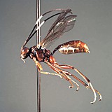
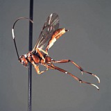
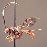
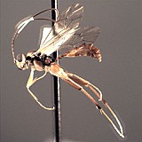
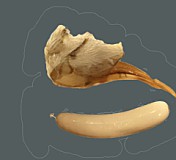
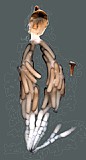
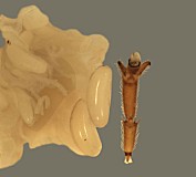
Clypeus highly variable among species and between sexes: varying from short and broad in some species to relatively tall and narrow in others; punctate, often coarsely so, especially apically, sometimes shagreened; ventral margin highly variable: sharp throughout in some species, thickened medially but sharp laterally in other species, when sharp, not distinctly impressed; shape of ventral margin either evenly convex, or broadly truncate medially and angled upwards laterally, or weakly concave in the middle of the truncate portion; also varying from sharp to blunt throughout, often blunt medially, sharp laterally; epistomal sulcus narrow, distinct, often sharply impressed, more rarely indistinct, sometimes varying between sexes; clypeus in profile flat to weakly bulging. Inner eye margins parallel. Malar space varying from slightly less than 0.5 times basal width of mandible to about 1.0 times basal width of mandible; malar sulcus absent. Mandible (Fig. 3) strongly curved, tapering from base to near middle then parallel-sided to weakly expanding from middle to apex; ventral tooth usually a little longer than dorsal tooth; ventral margin distinctly carinate. Maxillary palp shorter than height of head, less commonly equal to height of head; antenna (Figs 1, 3) usually slightly longer than body, first flagellomere moderate in length, typically as in Fig. 1 and 3. Ocelli small to moderate in size, diameter of lateral ocellus usually less than distance from lateral ocellus to eye. Hypostomal carina meeting occipital carina slightly but distinctly above base of mandible; occipital carina complete dorsally. Epomia sometimes absent or indistinct but present and distinct in at least three of the species examined. Epicnemial carina usually (95% of specimens examined) not reaching anterior margin of mesopleuron. Notaulus present usually as a weak impression on anterior declivity (the depression more rarely deeply impressed), weaker and indistinct on disk in some species, extending posteriorad level of tegula in other species though only rarely (one of the species examined) distinctly impressed. Groove between propodeum and metapleuron shallow, v-shaped, not u-shaped as in pionines; pleural carina present, usually strongly elevated; median longitudinal carinae very well-developed, forming flask-shaped median section with petiolar area broad and often delimited by median portion of posterior transverse carina; areola confluent with basal median area; lateral longitudinal carina weaker than median longitudinal carina, extending at least to spiracle from posterior margin and sometimes complete to anterior margin; posterior transverse carina complete in some species, completely absent in a few species; anterior transverse carina absent. Legs with apical margin of mid tibia either not expanded into a tooth or expanded into a tooth that is a little more rounded and not quite as well-developed as that of fore leg; apical comb on posterior side of hind tibia absent; posterior hind tibial spur 0.35-0.5 times length of long hind basitarsus (Figs 1-3); tarsal claws pectinate at extreme base in those species where the base was visible; fifth tarsomere of hing leg normal, not unusually elongate (relative to fourth) (Figs 1-3). Fore wing with areolet either present or absent, usually fairly consistent within a species; stigma moderately narrow as in Fig. 2, Rs+2r usually arising at or very slightly distad midpoint. Hind wing (Fig. 1) with first abscissa of CU1 either a little longer than, a little shorter than or equal in length to 1cu-a. T1 varying from relatively slender to somewhat broader: very gradually to more strongly expanding posteriorly; ventral margin usually weakly curved in profile; dorsal carinae absent or nearly so in the species examined; basal depression at dorsal tendon attachment varying absent or nearly so; dorsal-lateral carina varying from absent or nearly so in a few species to complete between spiracle and apex of T1 in some species; glymma absent. S1 varying among species: almost extending to level of spiracle in some species, extending to spiracle in others, and extending slightly posteriorad spiracle in some species. T2 thyridium not evident in species examined. Laterotergites of T2 and T3 separated by creases from median tergite. Ovipositor and sheath (Figs 1-3) downcurved or straight and posteriorly directed; ovipositor with distinct dorsal, subapical notch (Fig. 5); sheathes distinctive: outer surface of sheathes somewhat bulging, giving a slightly tubular appearance, usually broadest subbasally, truncate or nearly so at apex; hypopygium with distinctive pattern of short, erect setae. Male parameres elongate, extending distinctly beyond aedeagus in most individuals examined.

This description is based largely on about nine species in the Texas A&M University Collection and modified extensively from Townes (1970).

18453_mximage
1. Euryproctus habitus fema...
18452_mximage
2. Euryproctus habitus fema...
18454_mximage
3. Euryproctus habitus fema...
18455_mximage
4. Euryproctus habitus male...
6675_mximage
5. Euryproctusegg and oviposit...
6676_mximage
6. Euryproctu...
5969_mximage
7. Euryproctus egg and fore le...
 
Distribution
No referenced distribution records have been added to the database for this OTU.
Biology / Hosts
The eggs of at least one species (from central Texas) are large (Figs. 5-7 in description section), among the largest known for Ctenopelmatini (Cummins et al. 2011).
Map

There are no specimens currently determined for this OTU, or those specimens determined for this OTU are not yet mappable.

Acknowledgements
This page was assembled by Bob Wharton as part of a larger collaborative effort on the genera of Ctenopelmatinae. Page last updated April, 2015.

This work would not have been possible without the groundwork provided by Ian Gauld’s study of the Australian and Costa Rican faunas, and we are particularly grateful for his assistance in many aspects of this study. We thank David Wahl of the American Entomological Institute, Gavin Broad of The Natural History Museum, London, and Andy Bennett of the Canadian National Collection for extended loans of the material used for this study. We also thank David Wahl for useful feedback throughout our study, Anthony Cognato for specimens from Michigan, and Dimitry Kasparyan for a greater understanding of genera such as this one. Matt Yoder provided considerable assistance with databasing issues, and our use of PURLs (http://purl.oclc.org) in this regard follows the example of their use in publications by Norm Johnson. Heather Cummins, Caitlin Nessner, and Danielle Restuccia graciously assisted us with image processing, formatting, and literature retrieval. This study was supported by the National Science Foundation’s PEET program under Grant No. DEB 0328922 and associated REU supplement nos DEB 0723663, and 0923134.

This material is based upon work at Texas A&M University supported by the National Science Foundation under Grant Number DEB 0328922 with REU supplements DEB 0723663 and 0923134. Any opinions, findings, and conclusions or recommendations expressed in this material are those of the author(s) and do not necessarily reflect the views of the National Science Foundation.