Syndipnus Foerster, 1868

Taxonomic History / Nomenclature
Syndipnus Foerster, 1869: 209. Type species: Euryproctus (Syndipnus) macrocerus Thomson, 1883. Designated by Viereck (1914: 141) from among many species first included by Thomson (1883: 928).

Polypystis Foerster, 1869: 208. Type species: Tryphon lateralis Gravenhorst. Designated by Viereck (1914: 121) from among the species first included by Roman (1909: 315).

Tlemon Foerster, 1869: 209. Type species: Tlemon delicatus Ashmead, 1902. Subsequent inclusion by Ashmead (1902: 216). Monobasic. Synonymized by Townes (1945: 525)

Dicksonia Holmgren, 1880: 11. Type species: Dicksonia arctica Holmgren. Monobasic.

Neastus Holmgren, 1883: 154. Type apecies: Neastus laeviceps Holmgren, 1883: 154. Monobasic. Synonymized by Roman (1910).

Anaglymmus Roman, 1914. Type species: Anaglymmus incisus Roman, 1914. Designated by Viereck (1921). Synonymized by Townes (1945: 525).

The above synonymy follows Townes (1970: 131). Anaglymmus was included as a synonym of Synodites by Yu and Horstmann (1997). In Yu et al. (2012), the genus is listed as a synonym of Synodites, but the two originally included species are placed in Syndipnus. Townes (1970: 135) treats Anaglymmus as a synonym of Syndipnus.

Remarks
31 valid species were included by Yu et al. (2012).
Diagnosis and Relationships
Syndipnus is presently characterized largely on the basis of the fused T2+T3. T2 also bears a small, distinct tubercle mid-laterally. In habitus and several specific features, they resemble species in Synomelix, Pantorhaestes, Synodites, and Hypamblys. The absence of a fore wing areolet is useful for separating Hypamblys from Synomelix and Pantorhaestes.

There remains some doubt about the placement of several species in Syndipnus vs. Synodites and Hypamblys. Part of the issue is the placement of species in which the apical margin of the clypeus is neither entirely blunt nor entirely sharp. The second issue is the utility of the metasomal character used by Townes (1970: 129) to separate Syndipnus (with T2+3 fusion) from Hypamblys and other genera that have T2 and T3 clearly separate and overlapping. Aubert (2000) specifically states that the fused tergite character used by Townes does not work for some of the European species and Aubert says that Syndipnus is better characterized by the presence of warts on T2. Aubert also notes that Syndipnus is very difficult to separate from Hypamblys.

Description
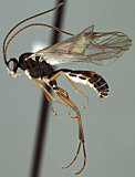
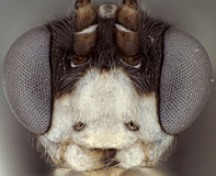
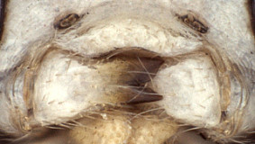
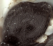
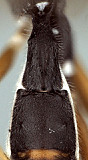
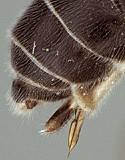
Clypeus (Fig. 2) usually narrow, with surface finely punctate; blunt, thickened at least medially, usually fairly sharp laterally; ventral margin varying from evenly but weakly convex to somewhat truncate medially and angled upwards laterally as in Fig. 2; epistomal sulcus narrow, distinct, often sharply impressed; clypeus in profile weakly bulging. Inner eye margins parallel. Malar space (Fig. 2) varying from about 0.5 to1.0 times basal width of mandible; malar sulcus absent. Mandible (Fig. 3) broader and tapering over basal 0.5, apical 0.5 parallel-sided; dorsal tooth usually broader and either equal in length to ventral tooth or very slightly longer; ventral margin distinctly carinate. Maxillary palp shorter than or equal to height of head; antenna (Fig. 1) about equal in length to body, first flagellomere usually short relative to species in genera such as Mesoleptidea and 
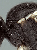
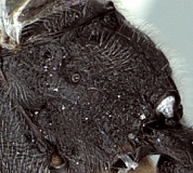
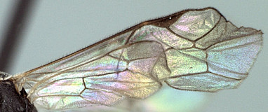
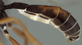
Hadrodactylus. Ocelli small to moderate in size, diameter of lateral ocellus usually less than distance from lateral ocellus to eye. Hypostomal carina meeting occipital carina distinctly above base of mandible; occipital carina complete dorsally. Epomia present, though sometimes weak and somewhat obscured by adjacent sculpture. Epomia absent or obscured by surrounding sculpture. Epicnemial carina usually reaching anterior margin of mesopleuron, but not in several males of one of the four species examined. Notaulus (Fig. 4) present usually as a distinct, sculptured impression on anterior declivity, becoming a little weaker and more shallow on disk, but converging posteriorly into shallow median depression at posterior margin of mesoscutum. Groove between propodeum and metapleuron absent to very weakly indicated, not u-shaped as in pionines; pleural carina present, but all carinae often obscured by dense propodeal sculpture in the species examined (Fig. 6); median longitudinal carinae, when visible, forming broad petiolar area posteriorly, narrowing anteriorly. Legs (Fig. 1) with apical margin of mid tibia expanded into a tooth that is not quite as well-developed as that of fore leg; apical comb on posterior side of hind tibia weakly developed; posterior hind tibial spur 0.4-0.55 times length of hind basitarsus; tarsal claws not pectinate; fifth tarsomere of hing leg normal, not unusually elongate (relative to fourth) (Fig. 1). Fore wing (Fig. 7) with areolet absent; stigma moderately broad, Rs+2r arising at or very near midpoint. Hind wing (Fig. 7) with first abscissa of CU1 usually about equal in length to 1cu-a, distinctly longer in at least one species. T1 varying from relatively slender (Figs 8, 9) to somewhat broader: very gradually to more strongly expanding posteriorly, respectively; ventral margin weakly curved in profile; dorsal carinae present, varying from low, barely extending to level of spiracles to distinctly elevated and extending at least 0,75 times length of T1 as in Fig. 9; basal depression at dorsal tendon attachment shallow; dorsal-lateral carina complete between spiracle and apex of T1; spiracle often placed basad midpoint of T1 (Fig. 8); glymma absent. S1 not extending to level of spiracle. T2 thyridium readily visible in at least some species. T2 and T3 at least partially fused (suture distinct, but the two tergite not movable relative to one another. Laterotergites of T2 and T3 separated by creases from median tergite. Ovipositor and sheath (Fig. 1) straight, ovipositor with distinct dorsal, subapical notch.

The above description is considerably modified from Townes (1970), and based on numerous specimens in the Texas A&M University collection.

10149_mximage
1. Syndipnus habitus...
10152_mximage
2.Syndipnus face
10153_mximage
3.Syndipnus mandibles
10155_mximage
4. Syndipnus mesosc...
10171_mximage
5.Syndipnus mesoscutum
10156_mximage
6.Syndipnus propodeum
10154_mximage
7.Syndipnus wings
10150_mximage
8.Syndipnus T1
10157_mximage
9. Syndipnus ...
10151_mximage
10. Syndipnus oviposi...
 
Distribution
No referenced distribution records have been added to the database for this OTU.
Biology / Hosts
Most host records are of Tenthredinidae and Diprionidae. Eller et al. (1989) recorded S. gaspesianus as the second most common parasitoid of Pikonema alaskensis (Rohwer) in their Minnesota (USA) sample site, and discussed diapause in the host and its parasitoids. Syndipnus rubiginosus was also reared from this host by Eller et al. (1989) and its sex pheromone identified (Eller et al. 1984).
Map

There are no specimens currently determined for this OTU, or those specimens determined for this OTU are not yet mappable.

Acknowledgements
This page was assembled by Bob Wharton as part of a larger collaborative effort on the genera of Ctenopelmatinae. Page last updated April, 2015.

This work would not have been possible without the groundwork provided by Ian Gauld’s study of the Australian and Costa Rican faunas, and we are particularly grateful for his assistance in many aspects of this study. We thank David Wahl of the American Entomological Institute, Gavin Broad of The Natural History Museum, London, and Andy Bennett of the Canadian National Collection for extended loans of the material used for this study. We also thank David Wahl for useful feedback throughout our study, Dimitry Kasparyan for a greater understanding of genera such as this one, and Dave Karlsson for access to material from the Swedish Malaise Trap Survey (trap 10, collection event 408; trap 50, collection event 1222). Matt Yoder provided considerable assistance with databasing issues, and our use of PURLs (http://purl.oclc.org) in this regard follows the example of their use in publications by Norm Johnson. Heather Cummins, Andrea Walker, Patricia Mullins, Caitlin Nessner, Amy James, Karl Roeder, Danielle Restuccia, and Cheryl Hyde graciously assisted us with image processing, formatting, and literature retrieval. This study was supported by the National Science Foundation’s PEET program under Grant No. DEB 0328922 and associated REU supplement nos DEB 0616851, 0723663, 0822676, 0923134, and 1026618.

This material is based upon work at Texas A&M University supported by the National Science Foundation under Grant Number DEB 0328922 with REU supplements DEB 0616851, 0723663, 0822676, 0923134, and 1026618.. Any opinions, findings, and conclusions or recommendations expressed in this material are those of the author(s) and do not necessarily reflect the views of the National Science Foundation.