Seleucus Holmgren, 1860
Taxonomic History / Nomenclature
Seleucus Holmgren, 1860: 111. Type species: Seleucus cuneiformis Holmgren, 1860. Monobasic.
Remarks
There are two valid species: Seleucus cuneiformis Holmgren, 1860 and Seleucus exareolatus Strobl, 1904 both Palaearctic.
Diagnosis and Relationships

Female Seleucus are readily recognized by the elongate and compressed metasoma, with the species much larger that those Saotis of similar habitus except in the case of Saotis seleuciformis (Kolarov). Males and females have T1 very narrow basally, abruptly expanded apically, with glymma absent. The medially carinate frons and very deeply cleft mandibular teeth also help in the recognition of this very distinctive taxon.
Description
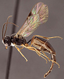
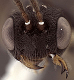
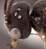
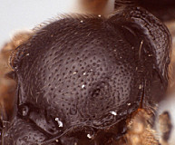
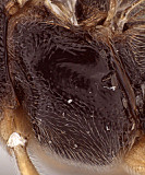
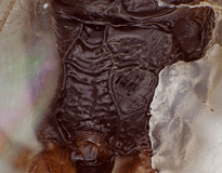
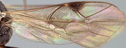
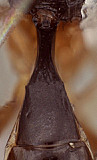
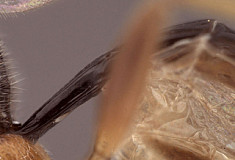
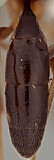
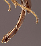
Clypeus (Fig. 2) strongly bulging, protruding in profile; ventral margin blunt, evenly convex; epistomal sulcus weakly separating clypeus from even more strongly bulging face. Malar space distinctly shorter than half basal width of mandible. Mandible (Figs 2, 3) very broad basally, with strong transverse, basal indentation, curved, strongly narrowing from base to middle, parallel-sided from midpoint to apex; ventral tooth usually about same size as dorsal tooth; ventral margin carinate; surface on basal half with diagonal ridge extending medially from dorso-basal corner. Frons with elevated carina along midline (Fig. 3). Female and male ocelli very small (Fig. 3), with maximum diameter of lateral ocellus distinctly shorter than distance between ocellus and eye. Female antennae much shorter than body (Fig. 1), first flagellomere not much longer than second. Hypostomal carina joining occipital carina a little above base of mandible; occipital carina complete. Epomia very well developed. Dorsal end of epicnemial carina extending towards anterior margin of mesopleuron but then extending parallel to it dorsally (Fig. 5), less commonly weakly separated from anterior margin; mesopleuron finely mat or polished, finely punctate. Notaulus essentially absent though punctation along notaular lines may be denser (Fig. 4). Small but distinct u-shaped groove present in lateral view between anterior end of lateral, longitudinal carina of propodeum and posterior margin of metathorax. Pleural carina very well-developed throughout; propodeal carinae complete: lateral and neduan longitudinal carinae well-developed throughout, fused posteriorly to form part of lateral margin of petiolar area; anterior and posterior transverse carinae also well-developed and complete except posterior transverse carinae sometimes indistinct among series of transverse ridges within petiolar area + areola (Fig. 6); when distinct, petiolar area and areola broad and well delineated, basal median area distinctly smaller. Legs with apical comb on posterior side of hind tibia present, short, moderately dense; hind tibial spurs slender (Fig. 1), longest spur about 0.4-0.5 times length of hind basitarsus; tarsal claws simple. Fore wing (Fig. 7) with areolet either present or absent (present in single female examined, absent in single male examined); stigma short, broad, Rs+2r arising from or near midpoint. Hind wing (Fig. 7) with first abscissa of CU1 longer than 1cu-a. T1 (Figs 7, 8) long, narrow, approximately parallel-sided over basal 0.45, abruptly broadening posteriorly as in Fig. 8; dorsal carinae varying from absent or not readily apparent to weakly elevated as low, short ridges medially; dorsal-lateral carina not apparent between spiracle and posterior margin; basal depression of dorsal tendon attachment absent; glymma absent. S1 not extending to level of spiracle, about 0.4-0.45 times length of T1. T2 thyridium absent; T2 and T3 exceptionally long, both in male (Fig. 10) and female (Fig. 1); laterotergites of T2 and T3 completely separated by creases in males, T2 separated by crease to level of spiracle in female, remainder of T2 and all of T3 pendulous; T3 of female with posterior margin narrow and deeply excised. Ovipositor (Figs 1, 11) short, upcurved, with deep subapical, dorsal notch; ovipositor sheath weakly clavate, but not as broad as in most Saotis. Apex of female metasoma as in Fig. 1: apical portion of female metasoma long and strongly compressed; female subgenital plate and at least two preceding sterna very long, desclerotized, and folded along midline.
This description is considerably modified from Townes (1971) and based on one female and one male in the Texas A&M University Collection.
Distribution
No referenced distribution records have been added to the database for this OTU.
Map
There are no specimens currently determined for this OTU, or those specimens determined for this OTU are not yet mappable.
Acknowledgements
This page was assembled by Bob Wharton as part of a larger collaborative effort on the genera of Ctenopelmatinae. Page last updated May, 2015.
This work would not have been possible without the groundwork provided by Ian Gauld’s study of the Australian and Costa Rican faunas, and we are particularly grateful for his assistance in many aspects of this study. We also thank David Wahl of the American Entomological Institute and Andy Bennett of the Canadian National Collection for extended loans of the material used for this study and particularly Dmitry Kasparyan for discussion of generic limits of this and other ichneumonids. Matt Yoder provided considerable assistance with databasing issues, and our use of PURLs (http://purl.oclc.org) in this regard follows the example of their use in publications by Norm Johnson. Heather Cummins, Andrea Walker, Caitlin Nessner, Mika Cameron, and Cheryl Hyde graciously assisted with image processing, formatting, and literature retrieval. This study was supported by the National Science Foundation’s PEET program under Grant No. DEB 0328922 and associated REU supplement #s DEB 0723663, 0923134, and 1026618.
This material is based upon work at Texas A&M University supported by the National Science Foundation under Grant Number DEB 0328922 with REU supplements DEB 0723663, 0923134, and 1026618. Any opinions, findings, and conclusions or recommendations expressed in this material are those of the author(s) and do not necessarily reflect the views of the National Science Foundation.
