Diachasmimorpha norrbomi Wharton, 2012
Taxonomic History / Nomenclature
Diachasmimorpha norrbomi Wharton, 2012. In: Wharton et al. (2012): 36-37 (key), 45-48 (description).
Remarks
This species is named for Allen Norrbom, who reared many Opiinae from various fruit, stem, and flower-infesting tephritids in Mexico and Central America.
Size variation in this species is similar to that exhibited by D. martinalujai, with males dominating the small end of the range.
Diagnosis and Relationships

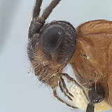
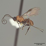
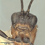
This species is similar in coloration to Diachasmimorpha hildagensis (Fischer) and Diachasmimorpha martinalujai Wharton but the ovipositor (with sheath 1.7-1.8 x longer than mesosoma) is slightly but distinctly shorter and the notaulus (Fig. 1) only rarely extends all the way to the anterior margin. The notaulus always reaches the anterior margin in the other two species (Fig. 2). Diachasmimorpha norrbomi is smaller and has a larger eye than Diachasmimorpha hildagensis (compare Figs 1 and 2), and 2RS tends to be longer (relative to 3Ra) in Diachasmimorpha norrbomi than in Diachasmimorpha hildagensis and Diachasmimorpha martinalujai.
Description
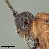
Female (Figs 1, 2). Head in dorsal view 1.25-1.30 x broader than mesoscutum, 1.80-1.85 x broader than face; eye in dorsal view 1.7-2.0 x longer than temple, temples not receding, but width at eyes greater than width at temples; eye in lateral view (Fig. 4) 2.1-2.9 x longer than temple. Facial midridge ending dorsally in short, very weak bifurcation between antennae. Frons irregularly rugulose along midline near bifurcation, otherwise polished, with moderately dense patch of decumbent, laterally-directed, white setae on either side of midline; bare on either side of ocellar field; width of ocellar field 1.0-1.2 x distance from ocellar field to eye. Face 1.80-1.95 x wider than high; uniformly setose (as in Fig. 3), distinctly punctate, punctures separated by at least 1 x their diameter. Malar sulcus deep, complete; malar space about 0.9-1.0 x basal width of mandible, 0.30-0.35 x eye height. Clypeus (Fig. 3) 2.8-3.2 x wider than high; very weakly convex, nearly flat. Occipital carina weak but distinct near base of mandible, short, extending dorsally to ventral margin of eye and often slightly beyond, not reaching mid eye height. Hypostomal carina extending as short but distinct flange below mandible. Antenna with 41-47 flagellomeres; first flagellomere 1.05-1.2 x longer than second; 1.8-2.0 x longer than wide.
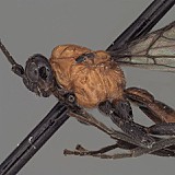
Mesosoma 1.35-1.45 x longer than high; 1.85-1.95 x longer than wide; 1.35-1.40 x higher than wide. Pronope deep, large, interrupting posterior crenulate groove middorsally; crenulae extending along dorsal 0.2 of pronotum laterally within narrow, shallow groove; groove not margined anteriorly by carina; anterior margin of pronotum laterally sinuate (Fig. 5), not abruptly excavated. Notauli (Figs 4, 5) deep anteriorly, gradually weakening posteriorly, extending posteriorly to level of tegula, not reaching long, narrow midpit, anterior end usually just short of and only rarely reaching anterior-lateral margin of scutum; mesoscutum usually without supra-marginal carina between base of notaulus and tegula, rarely with short, weak trace of a carina. Scuto-scutellar sulcus nearly rectangular, a little narrower medially; 4.2-4.8 x wider than midlength; crenulate-foveolate. Propodeum rugose, areola extending over posterior 0.8 but largely obscured by sculpture. Precoxal sulcus crenulate, widely separated from anterior margin of mesopleuron.
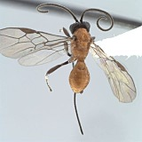
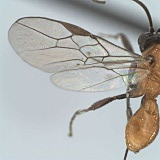
Wings (Fig. 6). Fore wing stigma short, broad, discrete distally, 3.15-3.30 x longer than wide; r1 arising from midlength of stigma; 1RS (excluding parastigma) 0.30-0.35 x length of 1M; m-cu postfurcal by 0.2-0.3 x length of m-cu; second submarginal cell distinctly converging distally; 2RS 1.0-1.2 x longer than 3RSa; 2CUa 1.6-1.8 x longer than 2cu-a; 1cu-a distad 1M by about 1.0 x its length.
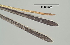
Metasoma not distinctly petiolate; head 1.6-1.9 x wider than apex of T1. T1 0.95-1.05 x as long as apical width; strongly diverging apically, with apex 2.0-2.5 x wider than base; surface smooth to weakly strigose posterior-medially, almost completed smooth laterally; dorsal carinae weakly converging, widely separated at posterior margin, strongly elevated over anterior 0.5, gradually weakening posteriorly; lateral carina weaker, extending distinctly ventrad spiracle, rounded and barely distinguishable posteriorad spiracle; spiracle at midlength of T1; dorsope absent but lateral and dorsal carinae elevated at junction, giving appearance of a slight depression; laterope deep; S1 very short, extending posteriorad to level of dorsal tendon attachment. T2 unsculptured, with sharp lateral margins. Ovipositor sheath (Figs 1, 2, 7) 1.7-1.8 x longer than mesosoma, setal pattern about as in D. martinalujai, with slightly greater density basally.
Color (Figs 1-4). Very similar to D. hildagensis. Meso- and metasoma orange, except tegula black; head dorsally dark brown to black except for small orange spot on vertex adjacent eye, lower occiput mostly yellow-orange, similar in color to broad band extending through epistomal sulcus, clypeus, lower gena (often), and mandibles; clypeus usually with narrow, transverse brown band, mandible with apical teeth dark, rarely with entire mandible brownish; legs black except extreme base and most or all of dorsal side of hind coxa orange, joint between femora and trochantelli reddish orange.
Male as in female except head in dorsal view 1.3-1.4 x broader than mesoscutum, 1.70-1.75 x broader than face; eye slightly smaller, in dorsal view eye 1.45-1.60 x longer than temple, in lateral view 1.9-2.4 x longer than temple; antenna with 41-43 flagellomeres, first flagellomere 0.95-1.2 x longer than second. Mesosoma slightly narrower, 1.95-2.05 x longer than wide; 1.4-1.5 x higher than wide; scuto-scutellar sulcus somewhat more variable in size, 4.0-5.5 x wider than midlength. Fore wing stigma 3.1-3.4 x longer than wide. T1 slightly smaller, head 1.9-2.2 x wider than apex of T1, T1 1.75-1.90 x wider at apex than at base.
Body length 3.3-4.3 mm, fore wing length 3.5-4.1 mm, mesosoma length 1.15-1.65 mm.
Distribution
Type locality. Mexico, Morelos, Parque Lago de Zempoala.
Paratypes: 27 females, 20 males, same data as holotype (TAMU, UNAM, USNM).
Other specimens examined (not paratypes): 1 female, 1 male, Mexico, D.F., Delegacion Tlapan, Fracc. Tlapuente, 19.ix.2003, M. Aluja #50, reared from fruit of Granadilla (TAMU).
In the original description of this species (Wharton et al. 2012), the type locality was incorrectly stated as being in the state of Mexico. The type locality is actually in the state of Morelos. The first line on the top label of the holotype (as given verbatim in the next section) and paratypes is therefore incorrect.
Distribution
No referenced distribution records have been added to the database for this OTU.
Biology / Hosts
The type series of Diachasmimorpha norrbomi was reared from Euphranta mexicana Norrbom infesting fruits of Ribes pringlei Rose (Norrbom 1993).
Two additional specimens that fit within the morphological limits of this species were reared from an unknown tephritid infesting Passiflora ligularis Juss.
Map
There are no specimens currently determined for this OTU, or those specimens determined for this OTU are not yet mappable.
Label data
Holotype. Female (UNAM), first label:
MEXICO: Mexico, Parque
Lag. de Zempoala, path
along L. Zempoala, 10-11.
VIII.1989, A.L.Norrbom
MEXICO: Mexico, Parque
Lag. de Zempoala, path
along L. Zempoala, 10-11.
VIII.1989, A.L.Norrbom
Second label:
reared ex. Euphranta
mexicana (Tephritidae)
ex. fruit of Ribes
pringlei Rose (89M13)
Acknowledgements
This page was prepared by Bob Wharton and is taken from a work on tephritid parasitoids published by Wharton, Lauren Ward and Istvan Miko (Wharton et al. 2012). Page last updated February, 2013.
We thank David Wahl (AEIC) and Kees van Achterberg (RMNH) for extended loans of holotypes, as well as Dominique Zimmermann and Manuela Vizek (NHMW), Gavin Broad (BMNH), Robert Kula and Paul Marsh (USNM), Jenö Papp (HNHM) and Henri Goulet (CNC) for assistance with loans of types and/or other material in their care. This work could not have been accomplished without the collecting efforts of several people, most notably Martin Aluja and his lab, Juan Rull, Al Norrbom, and Robert Jones. Imaging and plate assembly was considerably facilitated by Trent Hawkins, Karl Roeder, Cheryl Hyde, Patricia Mullins, and Sophia Daniels. Patricia Mullins and Matt Yoder provided assistance with databasing and the HAO. This work was supported in part by NSF DEB 0949027 and NSF/PEET DEB 0328922, with REU supplements 1213790 and 0616851 respectively (all to RAW). The HAO is funded by NSF DBI 0850223 to Andy Deans, formerly at North Carolina State University. RW prepared the descriptions of new species, LW assisted RW with opiine taxonomy and general manuscript preparation, and IM contributed the HAO linkages and a critical review of the morphological terms used throughout the published work.
