Lorenzopius van Achterberg and Salvo, 1997

Taxonomic History / Nomenclature
Lorenzopius van Achterberg and Salvo, 1997: 190-192. Type species: Lorenzopius calycomyzae van Achterberg and Salvo, 1997. Original designation.

Lorenzopius: Wharton et al. (2012): 32-34, 62-68 (review and redescription).

Type locality of calycomyzae: Cordoba, Argentina. Holotype female in National Museum of Natural History, Leiden.

Valid genus (van Achterberg and Salvo 1997)

Remarks
There are two distinct species groups within Lorenzopius: the calycomyzae species group containing the orginially included species Lorenzopius calycomyzae, Lorenzopius tubulatus, and Lorenzopius sanlorenzensis and a second group typified by Lorenzopius euryteniformis (Fischer). All have same basic wing venation and petiole. The precoxal sulcus is distinctly sculptured in the calycomyzae species group (Fig. 1) but the distinctly impressed sulcus is unsculptured or nearly so in the euryteniformis species group (Fig. 2). The smallest specimens of the calycomyzae species group examined during this study were slightly larger than the largest available specimens of the euryteniformis species group and perhaps as a consequence they tend to have slightly longer notauli and more sculpture bordering the supra-marginal carina extending from the base of the notaulus to the tegula. Most of the species we have examined from the euryteniformis species group have reduced propodeal sculpture with the median areola clearly visible.

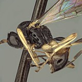
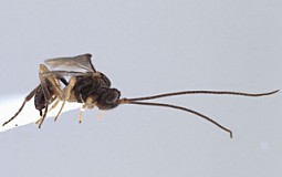
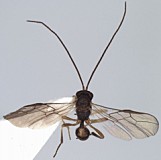
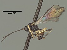
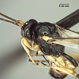
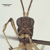
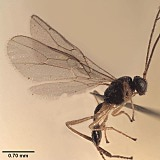
Lengthy descriptions (Fischer 1963, 1964, 1979, van Achterberg and Salvo 1997) and some redescriptions (Fischer 1977) are available for the described species of Lorenzopius and van Achterberg and Salvo (1997) provide a useful key to the species of the calycomyzae species group. See also the species pages for Lorenzopius calycomyzae, Lorenzopius tubulatus, and Lorenzopius euryteniformis.

19125_mximage
1. Lorenzopius tubulatus sc...
20570_mximage
2. Lorenzopius euryteniform...
 
Diagnosis and Relationships
Mandible (Fig. 4) distinctly narrowed from base to apex, without basal tooth or lobe ventrally. Labrum exposed (Figs 4, 8). Clypeus relatively flat, not distinctly protruding in profile; ventral margin sharp, truncate to weakly concave. Malar sulcus a sharp, weakly curved groove. Occipital carina broadly absent dorsally, present laterally; widely separated from hypostomal carina ventrally. First flagellomere longer than second. Propleuron ventral-laterally without oblique carina; pronotum dorsally without pronope or otherwise enlarged pit, posterior margin transversely rugulose. Notauli (Fig. 7) deep, narrow, well developed anteriorly, usually extending onto disc posteriorly; midpit present. Precoxal sulcus distinctly impressed. Propodeum (Figs 3, 10) with large median areola, posterior portion often obscured by rugose sculpture. Fore wing (Figs 6, 9) stigma long, narrow, parallel-sided, discrete posteriorly, r arising distinctly basad its midpoint but not from extreme base; m-cu entering base of second submarginal cell; second submarginal cell with 2RS shorter than 3RSb; 2CUb arising above middle of hind margin of first subdiscal cell. Dorsope and laterope of T1 absent (Figs 2, 7); S1 (Figs 1, 2) at least 0.7 x length of T1 in females, slightly shorter in males, apparently fused to T1; T1 long and narrow throughout Figs 3, 7); T2 and following terga unsculptured. Ovipositor tapering evenly to a fine point, without dorsal nodes or ridges.

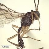
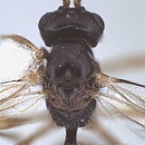
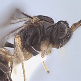
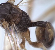
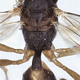
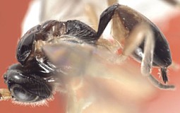
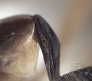
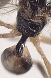
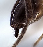
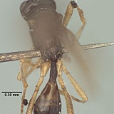
Lorenzopius and Tubiformopius are both characterized by having a tubular petiole with a long S1 which appears fused to T1 (Fig. 2). In the material available, S1 is longer in Lorenzopius than in Tubiformopius but there are more significant differences in the shape of the mandible, wing venation, and mesoscutal sculpture, as noted above in the section discussing genus group characters. Lorenzopius also shares many features with Eurytenes (Stigmatopoea), but the petiole is less tubular in the latter, with a distinctly shorter S1 that is clearly separated by membrane from T1.

Neither van Achterberg and Salvo (1997) nor Fischer (1998) mentioned the sternite in their descriptions, focusing instead on the tubular tergite, closed ventrally. What is most distinctive about Lorenzopius and Tubiformopius, however, is the length of S1 and its apparent fusion with T1. The presence of a prominent S1 is an unusual feature in the Opiinae and it is therefore not surprising that two genus group names have been proposed for species with this characteristic. The vast majority of opiine species have a very short basal sclerite, clearly separated by membrane from the tergite, but S1 is often difficult to see without dissection.

552_mximage
1. Lorenzopius calycomyzae holotype lateral...
551_mximage
2. Lorenzopius calycomyzae holo...
556_mximage
3. Lorenzopius ca...
553_mximage
4. Lorenzopius calycomyza...
19513_mximage
5. Lorenzopius calycomyz...
19120_mximage
6. Lorenzopius tubulatus holotype hab...
20514_mximage
7. Lorenzopius tubulatus ho...
20568_mximage
8. Lorenzopius euryteniform...
20572_mximage
9. Lorenzopius euryteniform...
20582_mximage
10. Lorenzopius euryteniform...
 
Description
19460_mximage
1. Lorenzopius calycomyzae holotype lateral...
19459_mximage
2. Lorenzopius calycomyzae ...
19512_mximage
3. Lorenzopius calycomyza...
25215_mximage
4. Lorenzopius calycomyzae ...
25216_mximage
5. Lorenzopius calycomyzae ...
19516_mximage
6. Lorenzopius calycomyzae hol...
19518_mximage
7. Lorenzopius calycomyzae ...
 
Distribution
No referenced distribution records have been added to the database for this OTU.
Biology / Hosts
The type species of Lorenzopius was described from specimens reared from Calycomyza mikaniae Spencer, a leafminer in the family Agromyzidae.
Map

There are no specimens currently determined for this OTU, or those specimens determined for this OTU are not yet mappable.

Acknowledgements
This page was assembled by Bob Wharton as part of research supported by NSF DEB 0949027 and NSF/PEET DEB 0328922, with REU supplements 1213790 and 0723663 plus 1026618 respectively (all to Bob Wharton). This page is based on the publication by Wharton et al. (2012). Page last updated February, 2013. The material on this page is freely available, but should be acknowledged if used elsewhere.

Imaging was considerably facilitated by Trent Hawkins, Cheryl Hyde, and Lauren Ward. Patricia Mullins and Matt Yoder provided assistance with databasing. I am particularly grateful to Kees van Achterberg (National Museum of Natural History, Leiden), Max Fischer and Dominique Zimmermann (Naturhistorisches Museum Wien), and David Wahl (American Entomological Institute, Gainesville) for extended loans of type specimens in their care. Ana DalMolin provided notes on the holotype of L. sanlorenzensis during her visit to the Hungarian Natural History Museum in Budapest.

This material is based upon work at Texas A&M University supported by the National Science Foundation under Grant Numbers DEB 0949027 and DEB 0328922 and associated REU supplements 1213790 and 0723663 plus 1026618. Any opinions, findings, and conclusions or recommendations expressed in this material are those of the author(s) and do not necessarily reflect the views of the National Science Foundation.