Metopius Panzer, 1806

Taxonomic History / Nomenclature
Metopius Panzer, 1806

Townes and Townes (1959) and Townes (1971) recognized seven subgenera. In addition to the subgenus Metopius, these are:
Peltastes Illiger, 1807 [= Tylopius Townes and Townes, 1959]
Peltocaris Thomson, 1887
Cultrarius Davis, 1897
Ceratopius Clement, 1927
Peltales Townes and Townes, 1959
Ethimetopius Benoit, 1961

Gauld and Sithole (2002)

abandoned the use of subgenera in their work on Costa Rican Metopius, preferring the use of species groups on practical and philosophical grounds. They treat subgenera as “nomenclatural encumbrances” and noted that while most of the subgenera of Metopius are distinctive, based on single autapomorphies, the recognition of some of these renders others paraphyletic (though they failed to provide any details).

Remarks
Nearly 150 valid species of Metopius were known as of 2011 (Yu et al. 2012). Of these, 49 are included in Metopius (Metopius), 34 in Peltastes, 25 in Ceratopius, 13 in Peltales, 12 in Cultrarius, 10 in Peltocaris, and 4 in Ethimetopius. Horstmann (2001) revised the Fabrician types of Ichneumon and concluded that Peltastes Illiger, 1807 was a senior synonym of Tylopius Townes and Townes, 1959. Yu et al. (2012) accordingly transferred all species previously placed in Tylopius to Peltastes. Horstmann (2001) noted that the type series of Ichneumon nectatorius Fabricius, 1773 consisted of three different species, and his lectotype selection resulted in movement of Peltastes from a junior synonym of Metopius s.s. to a senior synonym of Tylopius. Yu et al. (2012) listed the species described by Gauld and Sithole (2002) under the subgenus Metopius, but Gauld and Sithole specifically stated that their Metopius errantius species group was equivalent to Peltales even though they did not recognize subgenera.
Diagnosis and Relationships
Relationships of Metopius to other Metopiinae
In our analyses of relationships among metopiine genera (see Metopiinae page), we were unable to confirm the sister group relationship between Acerataspis and Metopius found by Quicke et al. (2009), despite the fact that this relationship was recovered in both the gap informative and gap uninformative analyses of Quicke et al. Our failure to recover this relationship was both surprising as well as disappointing because there is some morphological support for a Metopius + Acerataspis relationship. The most obvious example is the posteriorly protruding, flange-like lateral carinae of the scutellum, which is nearly identical in the species of these two genera. Strong lateral carinae are also present in Pseudometopius and Hemimetopius, but not quite so well developed. The closest approximation to the distinctive, shield-like face of Metopius, in our estimation, is found in Pseudometopius, proposed by Townes and Townes (1959) as the nearest relative to Acerataspis on morphological grounds. In our Bayesian analyses, Acerataspis forms an internal trichotomy, with other members of Group 1 contained within a second branch and a large clade anchored by Metopius forming the third branch. Sequence data for Pseudometopius, once obtained, may thus be the key to the resolution of the placement of Acerataspis, especially relative to Metopius. See additional comments on relationships among genera in the Metopiinae page.

Thus, three taxa within the Metopiinae (Hemimetopius, Pseudometopius, and Acerataspis) represent the closest intuitive sister taxa to Metopius. In addition to the features discussed in the previous paragraph, common character states shared with Metopius include presence of an areolet and interantennal process in Acerataspis and Pseudometopius. Additionally, Acerataspis shares presence of a single mesothoracic tibial spur. Hemimetopius is included for the shared characters of quadrate scutellum with strong lateral carinae, and apparently simple metathoracic tarsal claws. For our preliminary analysis of the subgenera of Metopius, all three taxa were coded, and each was analyzed separately with the Metopius taxa, and then analyzed as outgroup pairs. Exclusion of one or more of these as outgroup taxa lead to increased homoplasy and increased the number of equally parsimonious trees recovered. All three outgroups were therefore included in the subgeneric analysis whose results are given here to ensure that the number of trees recovered was not artificially high due to homoplasy within outgroups.

Assessment of Subgenera
We ran preliminary parsimony analyses on a relatively small dataset to explore relationships among the subgenera and to assess their monophyly. The character set that we used is given in the Description section below. Ingroup taxa for this preliminary analysis were as follows:
Metopius (Ceratopius) fuscipennis Wesmael, 1849
Metopius (Ceratopius) metallicus Michener, 1941
Metopius (Cultrarius) birkmani Brues, 1907
Metopius (Cultrarius) comptus Cresson, 1879
Metopius (Cultrarius) rufipes Cresson, 1875
Metopius (Cultrarius) xanthostigma Ashmead, 1890
Metopius (Ethimetopius) polyptichi Benoit, 1965
Metopius (Metopius) galbaneus Townes & Townes, 1959
Metopius (Metopius) insularis Seyrig, 1934
Metopius (Metopius) rufus Cameron, 1905
Metopius (Metopius) syriacus Clément, 1930
Metopius (Metopius) vittatus Townes & Townes, 1959
Metopius (Metopius) zuluanus Benoit, 1965
Metopius (Peltales) amenus Gauld & Sithole, 2002
Metopius (Peltales) notatus Townes & Townes, 1959
Metopius (Peltastes) bellus Cresson, 1879
Metopius (Peltastes) edwardsii Cresson, 1879
Metopius (Peltastes) leiopygus Foerster, 1850
Metopius (Peltocaris) croceicornis Thomson, 1887
Metopius (Peltocaris) dentatus Fabricius, 1779

In our preliminary parsimony analysis, Ceratopius, Cultrarius, Peltales, and Peltocaris were recovered as monophyletic in all 6 equally parsimonius solutions. The species of Peltastes formed a basal grade to a larger clade containing Ceratopius + Peltales, and Metopius was paraphyletic. Ethimetopius was represented in the consensus tree by a single taxon forming part of a large polytomy including all but the Cultrarius clade and two of the Metopius species.

Members of the subgenus Metopius are united as a subgenus by the presence of an occipital carina that extends below the foramen magnum and is thus complete or nearly so (Townes and Townes 1959). Nearctic species have two mandibular teeth, the lower tooth present as a small projection on the outer edge of the upper tooth (Townes and Townes 1959). Our analysis provides a specific example for the more general statement by Gauld and Sithole (2002) that recognition of some subgenera will render others paraphyletic. Metopius is the extreme case and our results strongly suggest that any effort to develop a classification in which all subgenera are monophyletic would necessitate dismantling Metopius s.s. into a series of smaller subgenera.

Peltocaris is strongly supported by the shape of the interantennal process, which extends forward onto the facial shield, a clypeus that slopes outwardly, with clypeal margin that lacks a distinct carina, and reduced propodeal sculpture.

Our results also support the monophyly of Cultrarius, based on the character states of the distally flattened antenna and short occipital carina used by Townes and Townes (1959). Townes and Townes (1959), in their catalog of Eastern Palearctic Ichneumonidae, tentatively placed M. turcestanicus Clément, 1930 in Cultrarius, but our examination of this species suggests that it lacks the diagnostic features of Cultrarius and should be placed elsewhere, leaving Cultrarius as a New World endemic. Similarly, Ceratopius is strongly supported as monophyletic by the same diagnostic feature used by Townes and Townes (1959): the small horn on the frons that is distinct from the interantennal process, as well as the shape of the interantennal process. The monophyly of Peltales is supported by the shape of the square to trapezoidal interantennal process with elevated margins. Gauld and Sithole (2002) provide additional diagnostic features.

Our results are decidedly preliminary as the coding of some characters proved to be more subjective than we had initially hoped, but is provided here to highlight where major problems lie in using a subgeneric classification for Metopius.

Description
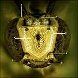
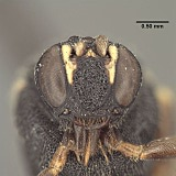
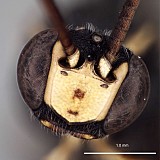
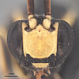
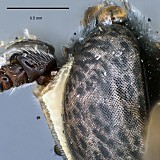
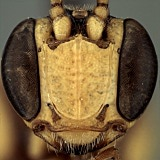
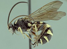
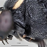
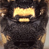
Metopius is readily characterized and separated from all other members of the Metopiinae by the distinctive shield-shaped carina that encompasses the face, forming a facial shield (Fig. 1, #3 and Figs 2-3). Additionally, the epipleurites (=lateral tergites) are always well-developed, there is a single tibial spur on the mesothoracic leg, and the first two metasomal tergites are fused.

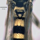
The species of Metopius are highly color-variable and this variation has been used extensively to delimit species and subspecies. The following characters and character states encompass much of the remaining morphological variation in Metopius and these features were used to explore relationships among the recognized subgenera, as detailed in the sections above.

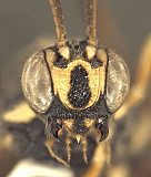
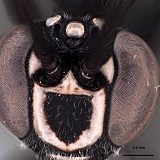
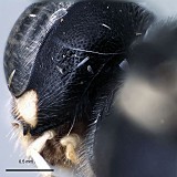
The interantennal process (#2 in Fig. 1) extends between the antennae, from the facial shield to the frons. It is present in all species of Metopius, exhibiting the following variation: Laterally compressed, with anterior portion extending forward onto the dorsal part of the shield as a ridge-like projection (Fig. 4); laterally compressed, at least dorsally, without anterior ridge (Fig. 5); anterior portion square to trapezoidal, concave, with raised marginal flanges (Fig. 6); anterior portion broadly triangular and flattened (Fig. 7); anterior portion broadly triangular, posterior portion concave, with raised marginal flanges (Fig. 8).

The frontal horn is a distinct projection physically separated from and posterior to the interantennal process. It is present in a few taxa, such as M. fuscipennis (Fig. 9) and M. metallicus, but absent in most or present but connected to the interantennal process by a distinctly elevated flange (as in M. birkmani).

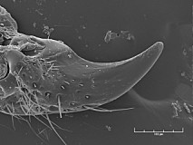
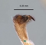
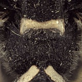
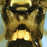
The lower or ventral tooth of the mandible may be either absent (Fig. 10) or present. When present, it may be represented by a small, inwardly-directed projection not separated by a groove from the dorsal tooth; it may be small and inwardly-projecting, but separated by a distinct groove or indentation from the dorsal tooth (Fig. 11); or it may be larger: only slightly smaller than the dorsal tooth and in the same plane as the dorsal tooth.

The occipital carina is present dorsally, but variously absent ventrally: either relatively complete and extending well below the level of the middle of the foramen magnum (Fig. 12) or relatively incomplete and extending ventrally at most to the middle of the foramen magnum.

Clypeal margin: the clypeus and face are not distinctly separated and the clypeal margin (Fig. 1, # 6) thus appears to be the ventral margin of the face. The margin may be carinate, with the carinate margin weakly protruding (Fig. 13); the entire clypeus may be sloping outwardly so that it overhangs the base of the labrum (Fig. 14); or the margin may be impressed and not distinctly protruding (Fig. 15).

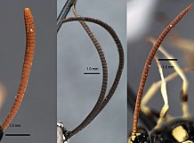
The flagellum is normally cylindrical to slightly flattened in the middle, with the flagellomeres distinctly tapering distally (Fig. 16, middle and right). In some species, however, the flagellomeres are more strongly flattened in the distal third (Fig. 16, left).

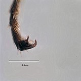
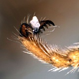
The tarsal claws of the pro- and mesothoracic legs may be either simple (Fig. 17) or pectinate (Fig. 18). In nearly all species with pectinate claws, the pectination is confined to the basal half and is often weak (Fig. 19).

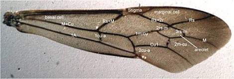
Cross-vein 2m-cu of the fore wing may have either 1 (Fig. 20) or 2 bullae. Unfortunately, this character is subject to some intraspecific variation. Removal of this character from our preliminary analyses resulted in fewer equally parsimonious trees.

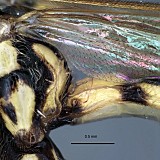
The fore wing color pattern varies as follows: Uniformly colored, either hyaline or variously yellow (Fig. 20); bicolored, with anterior 1/3 of the wing dark brown from base to apex, remainder hyaline or nearly so; or bicolored but darkening distally (Fig. 21), with dark yellow to brown coloration limited to the marginal cell and apex of wing, cells basal to stigma never darkened.

The epicnemial carina has three character states, with the first two somewhat subjective: Carina absent dorsally, ending just above the sternaulus and widely separated from the anterior and dorsal margins of the mesopleuron (Fig. 22); carina gradually turns towards anterior margin of mesopleuron with no distinct turn, ending parallel at a distance equal to or greater than the width of an ocellus (Fig. 23); carina turns sharply towards anterior margin of mesopleuron above the sternaulus before turning again and ending at or closely parallel to anterior margin of mesopleuron (Fig. 24), leaving a width less than the diameter of an ocellus.

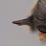
A tibial tooth is usually present as a narrow, distinctly pointed apical extension of the dorsal rim of the metathoracic leg, opposite the tibial spurs (Fig. 25), but is absent or nearly so and broadly rounded in some species.

The shape of the subalar prominence is considerably variable, and we attempted to capture this variability with the following character states: weakly convex; narrowly and more strongly convex; narrow and ridge-like.

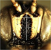
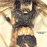
Fusion of the first and second metasomal tergites (T1 and T2) is characteristic of Metopius. Though fused, the suture is still visible as an impressed line, either continuously across the middle (that is, uninterrupted: Figs 26, 30) or the suture may be interrupted by a pair of longitudinal carinae from T1 (Fig. 27) that extend onto the base of T2, obliterating the suture line medially. In outgroup taxa Acerataspis, Hemimetopius, and Pseudometopius, there is no fusion of T1 and T2.

The male clasper is either evenly convex or depressed apical-ventrally, resulting in a lateral ridge.

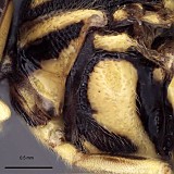
Propodeal carinae are variously reduced or absent and the following characters states address this variation: greatly reduced, with no closed cells, anterior transverse carina (ATC) and posterior transverse carina (PTC) absent, short remnants of median longitudinal carinae anterior and lateral longitudinal carinae posteriorly usually present; pleural carina present (Fig. 28); moderately reduced: either ATC present laterally, but median longitudinal carinae mostly absent (Fig. 29); OR ATC largely absent but median basal area + areola distinct; nearly complete areolation, with closed cells anteriorly and laterally, but with most of PTC absent and basal area fused with areola, closed posteriorly by short median section of PTC, with median section of ATC absent (Figs 30, 31); completely areolate, ATC, lateromedian longitudinal carina, lateral longitudinal carina, and PTC present as strongly elevated carinae (condition in some outgroups, but not in any Metopius we have seen.

23517_mximage
1. Metopius head, anterior ...
6429_mximage
2. Metopius head, ante...
23521_mximage
3. Metopius head, anterior ...
24160_mximage
4. Metopius (Peltocarus) cr...
20621_mximage
5. Metopius (Cultrarius) ri...
23501_mximage
6. Metopius (Peltales) erra...
23514_mximage
7. Metopius (Peltastes) edw...
24168_mximage
8. Metopius (Ceratopius) me...
24303_mximage
9. Metopius (Ceratopius) fu...
5162_mximage
10. Metopius (Cultrarius) comptus man...
20826_mximage
11. mandible with two teeth,...
24169_mximage
12. Pseudometopius head in p...
20799_mximage
13. Metopius (Cultrarius) bi...
24159_mximage
14. Metopius (Peltocarus) co...
23510_mximage
15. Metopius (Peltastes) bel...
24311_mximage
16. Left to Right: Metopius (Cultrariu...
23504_mximage
17. Metopius (Peltastes) pol...
20807_mximage
18.pectinate tarsal claws.
24173_mximage
19. Pseudometopius tarsal cl...
6425_mximage
20. Metopius habitus showing 1 bulla a...
23519_mximage
21. Metopius forewing showing bicol...
20859_mximage
22. Metopius (Cultrarius) xa...
24316_mximage
23. Metopius (Peltales) nota...
24325_mximage
24. Metopius (Peltastes) pol...
24320_mximage
25. Metopius metallicus tibi...
20617_mximage
26. Metopius (Cultrarius) pe...
24324_mximage
27. Metopius (Peltales) nota...
20796_mximage
28. Metopius (Peltastes) bas...
20821_mximage
29. Metopius (Metopius) krom...
23138_mximage
30. Metopius (Peltales) nota...
24318_mximage
31. Metopius (Peltales) nota...
 
Distribution
Metopius is worldwide in distribution. Members of the subgenus Metopius are found in the Holarctic, Oriental, and Australian Regions and members of the subgenus Ceratopius occur in the Palaearctic and Oriental Regions. Members of the subgenus Peltocaris are found throughout the Palaearctic Region and the known species of Ethimetopius are exclusively Afrotropical. Peltastes is Holarctic, Cultrarius is primarily Nearctic with one species recorded from the Palaearctic (but see comments under this subgenus in the diagnosis and relationships section above), and the species of Peltales are exclusively New World in distribution.
Distribution
No referenced distribution records have been added to the database for this OTU.
Biology / Hosts
The bicolored wing patterns of some species are thought to mimic the dorsal-longitudinal wing folding of eumenine wasps of the family Vespidae. Townes (1959) made several observations of Metopius behavior that he equated to eumenine mimicry.
Map

There are no specimens currently determined for this OTU, or those specimens determined for this OTU are not yet mappable.

Acknowledgements
This page was assembled by Bob Wharton and Danielle Restuccia. It contains freely available unpublished work by Wharton and Aubrey Colvin at Texas A&M University and was supported by the National Science Foundation under PEET grant # DEB 0328922 and associated REU supplement # 0723663. We thank David Wahl (American Entomological Institute), Andy Bennett, and Bob Zuparko (California Academy of Sciences) for extended loans of material used for this study. We are particularly grateful to Gavin Broad, who provided feedback on this webpage.

Some details of the unpublished analyses shown here are available from Wharton. Page last updated September, 2013.

This material is based upon work supported by the National Science Foundation under Grant Number DEB 0328922 and REU supplement number 0723663.
Any opinions, findings, and conclusions or recommendations expressed in this material are those of the author(s) and do not necessarily reflect the views of the National Science Foundation.