Neurogenia ocellaris Benoit, 1955
Taxonomic History / Nomenclature
Neurogenia ocellaris Benoit, 1955: 69, 72-73.
Diagnosis and Relationships

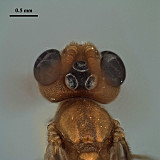
Ocelli exceptionally large in both sexes, though notably more so in male with length of lateral ocellus > 6 x the distance between lateral ocellus and eye. Spurious vein well-developed on M+Cu. Propodeal areola not delimited anteriorly, propodeal surface sculptured medially. Similar to both N. appendiculata and an undescribed species from Cameroon in possessing large ocelli, but differing from the former by the absence of a malar space and from the latter by the well-developed spurious vein.
Description
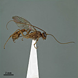
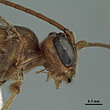
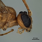
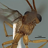
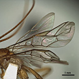
Mesosoma 2.2 mm; fore wing 5.8-5.9 mm. Antenna with 52 (female) and 46 [46-47 in original description] (male) flagellomeres. Frons coarsely granular, abruptly transitioning posteriorly at anterior margin of lateral ocellus to punctate, the punctures separated by about their diameter. Eye large, in lateral view 1.6 (female) and 2.0 (male) x longer than temple (Figs 2, 3). Ocelli exceptionally enlarged, maximum diameter of lateral ocellus 1.75 (female) and 3.55 (male) x distance from lateral ocellus to eye (male: Fig. 4); ocellar triangle 3.6 (female) and 6.9 (male) x wider than shortest distance between lateral ocellus and eye. Malar space barely indicated in female. Epomia well-developed, vertical. Mesoscutum punctate, punctures separated by about their own diameter laterally, becoming larger and denser posteromedially; notaulus weakly indicated on anterior declivity. Propodeal carinae nearly complete (Fig. 5): areola narrowing posteriorly, confluent with basal area anteriorly (basal transverse carina absent or very weak medially); propodeum rugulose medially, second lateral area weakly punctate in male, more heavily and densely punctate in female. Fore wing Rs+2r arising distinctly distad mid stigma, from about apical 0.4; M+Cu with spurious vein (Fig. 6) extending into basal cell more than half distance to anterior margin, bent or curved distally in its anterior 0.25-0.3, spurious vein distant from 1M by about 1.4 x length of 1cu-a; 1cu-a distinctly postfurcal; 1M weakly sinuate, nearly vertical for most of its length, slightly curving distally into parastigma; m-cu parallel to 1M basally, evenly curving distally; basal cell largely bare on either side of spurious vein. Hind wing cu-a long to very long, 3.5 (female) and 10.4 (male) x longer than 1st abscissa of Cu1. Metasoma with T1 length from spiracle to base 2.1-2.2 x longer than width at spiracles 4.0-4.2 x longer than minimum width, width at spiracle 1.9 x minimum width. Color: yellow-orange, including all flagellomeres, except ocellar field dark, mandibular teeth dark red; wings very faintly infumate.
This description and the associated images are based primarily on a male and female from Gambia in BMNH. The male matches the original description and notes on Benoit’s holotype (RMCA).
Distribution
This species was described from two males collected in Pobe, Dahomey (=Benin) (Repository: RMCA). We have seen two additional specimens from Gambia (BMNH), with the male perfectly matching the original description.
Distribution
No referenced distribution records have been added to the database for this OTU.
Biology and Behavior
Biology unknown.
Map
There are no specimens currently determined for this OTU, or those specimens determined for this OTU are not yet mappable.
Acknowledgements
This page was assembled by Bob Wharton as part of a larger collaborative effort on the genera of Ctenopelmatinae, and as part of a study of this genus prepared by Heather Hendrickson and Bob Wharton. The work is based on specimens in the Texas A&M University collection as well as material borrowed from China, MRAC, CNC, BMNH, and AEI. We are particularly grateful to Xue-xin Chen, David Wahl, Andy Bennett, Gavin Broad, and Eliane De Coninck for the loan of valuable material. This work would also not have been possible without the groundwork provided by Ian Gauld’s study of the Australian and Costa Rican faunas, and we are particularly grateful for his assistance in many aspects of this study. We thank David Wahl for useful feedback throughout our study and to Gavin Broad for exchange of information on Perilissini. Matt Yoder provided considerable assistance with databasing issues, and our use of PURLs (http://purl.oclc.org) in this regard follows the example of their use in publications by Norm Johnson. Lauren Ward graciously assisted with image capture, processing, and formatting. Page last updated December, 2014.
This material is based upon work supported by the National Science Foundation under Grant Number DEB 0328922 with REU supplements DEB 0723663 and number 1026618.
Any opinions, findings, and conclusions or recommendations expressed in this material are those of the author(s) and do not necessarily reflect the views of the National Science Foundation.
