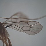
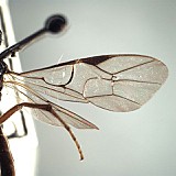
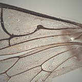
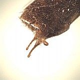
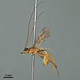
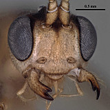
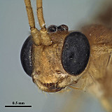
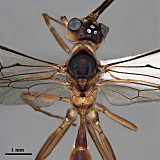
Clypeus somewhat elliptic, weakly separated from face by broad, shallow, indistinct epistomal sulcus (Figs 3-5); nearly flat in profile; apical margin thickened and weakly convex to more or less truncate over medial 0.4-0.6, outer margin of thickened portion with an irregular row of low, rounded bumps (Figs 3, 4); apical margin thin and recurved laterally (Figs 3, 4). Anterior tentorial pits large, elliptical, with narrow, distinctly impressed groove from lateral margin of pit to base of mandible. Mandible (Figs 3-5) distinctly longer than broad; outer face flattened basally, curved apically with apical portion somewhat reflected so that the dorsal margin is angled towards the clypeus relative to the ventral margin; ventral tooth about twice length of dorsal tooth, both well-developed. Malar space (Figs 3, 4) short, sometimes absent, interspecifically variable because eye in male often larger than eye in female (Figs 6, 7); when malar space present, malar sulcus absent. Occipital carina complete dorsally, extending ventrally to mandible as a well-defined ridge; hypostomal carina appears widely separated from occipital carina at base of mandible, but actually curves along base of head capsule to join occipital carina usually at ventral condyle of mandible (Fig. 2). Antenna at least as long as body, often distinctly longer; flagellomeres distinctly longer than wide; first flagellomere 1.2-1.35 times longer than second, with distinct patch of tyloids at extreme base on outer side. Ocelli variable in size, with maximum diameter of lateral ocellus less than, equal to, or much greater than distance between ocellus and eye margin. Frons weakly depressed medially between bases of antennae and median ocellus, without median vertical projections. Pronotum dorsally with broad, transverse groove; pronotal collar with posterior margin indented mid-dorsally due to strongly protruding, chevron-shaped anterior margin of mesoscutum; pronotum laterally with anterior margin thin, sharp, and straight or nearly so, not obviously sinuate; not or only very weakly inflated below mesoscutal suture; epomia varying from long and distinct throughout (Fig. 8) to short, not extending in either direction much beyond vertical groove (Fig. 7). Notaulus at most represented by a faint impression anteriorly. Scutellum carinately margined basally, but usually only at extreme base. Epicnemial carina well-developed, dorsal end distant from and not curved towards anterior margin of mesopleuron; mesopleuron with groove extending from mesopleural fovea to dorsal end of epicnemial carina; subtegular ridge thin, sharp throughout, protruding at a right angle from mesopleuron; posterior margin of mesopleuron crenulate above and below mesopleural fovea. Metapleuron as in Fig. 14. Propodeum (Figs 14, 15) completely carinate in most species; lateral longitudinal carina complete from apex to base; lateromedian carina often complete but occasionally effaced or greatly weakened medially and/or anteriorly, in which case areola incomplete; median apical area often with an additional median carina; basal and apical transverse carinae usually well defined (basal transverse carina absent medially in a few species), extending between fully developed pleural carinae; surface of propodeum varying from rugulose to polished and unsculptured; trough along anterior margin wide, narrowing slightly but remaining deep and distinct between apices of median and lateral longitudinal carinae (anterior rim of propodeum thus distinctly separate from posterior rim of metanotum in this area). Legs, including hind femur, slender; hind tibial spurs unequal, longest spur slender, 0.25-0.45 times length of basitarsus; apex of hind tibia posteriorly with dense comb of setae; apex of mid tibia lacking distinct tooth characteristic of anterior tibia; tarsal claws sparsely but distinctly pectinate. Fore wing stigma wedge-shaped to nearly hemi-elliptical, RS+2r arising near midpoint (position varying among species); M+CU near its middle either tuberculate (with a thickened bulge) or with a short, anteriorly-directed spurious vein; basal vein (1M) either evenly bowed distally, and usually thickened, or straight to weakly sinuate, never evenly bowed basally as in most other ctenopelmatine genera; areolet nearly always (95%) present, petiolate, with 3rs-m bowed; second recurrent vein (2m-cu) with a single bulla, often occupying most of anterior half of vein; 1cu-a usually postfurcal, more rarely antefurcal; 1A straight throughout; 2A rarely a discrete vein, usually represented only by a broad basal smudge. Hind wing M+CU weakly arched; first abscissa of CU1 commonly very distinctly shorter than 1cu-a; 1cu-a reclivous, rarely vertical when very short. T1 with spiracle at midlength; heavily sclerotized portion of sternum extending to end of deepest portion of glymma but not to spiracle, weakly separated from but not fused to tergum; glymma deep and broad within basal third of petiole, the two sides of the pit separated by a thin, translucent sclerite, glymma tapering anteriorly to base and posteriorly to ventral margin, the groove subtended by a carina terminating posteriorly about level of spiracle; extreme base of petiole flat dorsally, without depression; dorsal-lateral margin very sharp from base to middle of dorsal margin of glymma, from there gradually rounding to spiracle; dorsal-lateral carina absent posteriorad spiracle. T2 and T3 not or only indistinctly punctate, otherwise unsculptured; T2 thyridium and gastrocoeli absent; epipleura of T2 and most of T3 usually separated by a sharp crease from the median tergite. Cercus about 2-3 times longer than wide. Subgenital plates of male and female with apical margins simple. Dorsal notch of ovipositor nearly always a broad, weak depression that is somewhat indistinct (Fig. 19); only very rarely with a distinct notch. Male parameres long (Fig. 17), often very long and narrow (Fig. 18); aedeagus rounded, knob-like apically.
This description is greatly expanded from Townes (1970), and is based on both described and undescribed species, mostly from AEI, BMNH, CNC, and TAMU. An earlier version from this website was used by Reshchikov et al. 1914.
2.
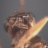
juxtaposition of occipit...
juxtaposition of occipital and hypostomal carinae
↰
↴
5.
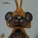
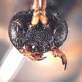
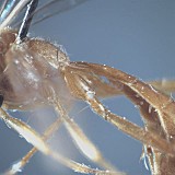
N. hunanensis face and c...
N. hunanensis face and clypeus
↰
↴
6.
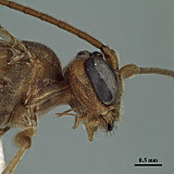
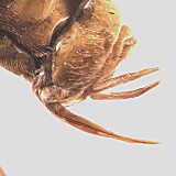
Neurogenia ocellaris fem...
Neurogenia ocellaris female
↰
↴
7.
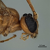
Neurogenia ocellaris mal...
Neurogenia ocellaris male
↰
↴
8.
Neurogenia cubitalis lon...
Neurogenia cubitalis long epomia
↰
↴
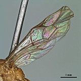
12.
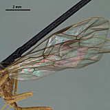
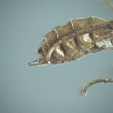
N. tuberculata fore wing...
N. tuberculata fore wing
↰
↴
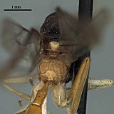
14.
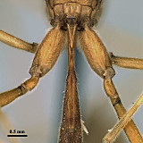
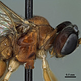
lateral view, mesosoma a...
lateral view, mesosoma and T1
↰
↴
15.
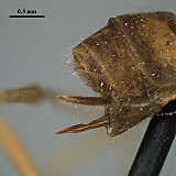
kamapura propodeum...
kamapura propodeum
↰
↴
16.T1 showing deep glymma
18.
N. shennongjiaensis male...
N. shennongjiaensis male genitalia
↰
↴