Rhinotorus Foerster, 1869

Taxonomic History / Nomenclature
Rhinotorus Foerster, 1869: 211. Type species: Spudaea longicornis Schmiedeknecht, 1914. Designated by Perkins (1962: 450).

Spudaea Foerster, 1869: 211. Type species: Tryphon leucostomus Gravenhorst, 1829. Subsequent designation by Viereck (1914: 136) from among three species first included in Spudaeus by Thomson (1883: 932). Synonymized by Perkins (1962).

Spudaeus Thomson, 1883. Unjustified emendation.

Prospudaea Hincks, 1944. New name for Spudaea Foerster, 1860 not Spudaea Snellen von Vollenhoven, 1867.

Remarks
The following valid species were included in this Holarctic genus by Yu et al. (2012).

Rhinotorus alpinus (Roman, 1909)
Rhinotorus brachycerus Kasparyan and Kopelke, 2009
Rhinotorus clypeatus (Brischke, 1888)
Rhinotorus compactor (Thunberg, 1822)
Rhinotorus latvicus (Ozols, 1928)
Rhinotorus leucostomus (Gravenhorst, 1892)
Rhinotorus longicornis (Schmiedeknecht, 1914)
Rhinotorus mesocastanus (Thomson, 1893)
Rhinotorus nasutus (Gravenhorst, 1829)
Rhinotorus ovalis (Davis, 1897)
Rhinotorus similis (Brischke, 1892)
Rhinotorus umbrarum (Holmgren, 1857)

Diagnosis and Relationships
Rhinotorus can be recognized by the transverse, subapical impressions on T2 (and usually on T1 and T3). The impressions are shallow, and not always easy to distinguish among the heavy sculpture. Additionally, the clypeal margins are usually sharp at least laterally and the claws are variously pectinate.
Description
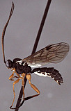
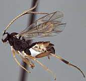
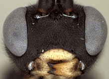
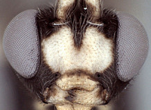
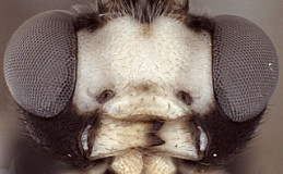
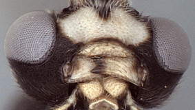
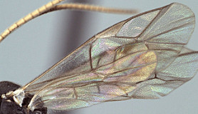
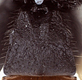
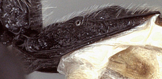
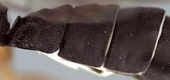
Frons without median horn or elevated carina. Clypeus small (Figs 3-6) narrow and short, with transverse, subapical bulge that is well developed and overlapping apical margin medially in some species (Fig. 3) but nearly absent in others; ventral margin otherwise sharp throughout, truncate (Fig. 3) or weakly concave (Fig. 4) medially, with sharp lateral margins distinctly angled dorsally; epistomal sulcus distinct in some species, indistinct in others. Malar space varying from about 0.4 to 0.9 times basal width of mandible. Mandible (Figs 3-6) curved, gradually narrowing from base to apex, ventral tooth varying from about equal in length to somewhat shorter than dorsal tooth and usually narrower than dorsal tooth; when mandibles closed, clypeus sometimes obscures part of mandible and mandible sometimes obscures part of clypeus. inner eye margins parallel to very weakly converging ventrally. Ocelli small, with maximum diameter of lateral ocellus distinctly shorter than distance between ocellus and eye. Female antenna equal to or longer than body. male antenna usually shorter than body; first flagellomere long and slender in one species examined, but shorter in others (as in Figs 1, 2). Hypostomal carina joining occipital carina well above base of mandible; occipital carina complete. Epomia absent. Dorsal end of epicnemial carina extending to anterior margin of mesopleuron in all specimens examined; mesopleuron finely to coarsely mat ventrally, nearly rugulose in some species. Notaulus sharply impressed on anterior declivity (though not quite reaching anterior margin in one of the species examined), distinctly impressed on disk at least to level of tegula. Pleural carina complete, well-developed; propodeum (Figs. 7, 8) with lateral longitudinal carina well-developed, complete or nearly so though usually weaker anteriorly; median longitudinal carinae forming a broad, rounded petiolar area then extending anteriorly as sharp, more narrowly spaced ridges; short, broad petiolar area usually with median longitudinal carina (Fig. 7) and separated by transverse carina from combined areola plus basal median area; posterior transverse carina present across middle, extending to lateral longitudinal carina; anterior transverse carina completely absent. Hind tibia with apical comb on posterior side absent or poorly developed; hind tibial spurs long, slender, spurs unequal in size, longest spur 0.5-0.8 times length of hind basitarsus; all tarsal claws usually pectinate, but extent and development of pectination varies among species. Fore wing (Fig. 9) with areolet absent; stigma not somewhat narrow, with Rs+2r arising from or a little basad midpoint. Hind wing (Fig 9) with first abscissa of CU1 longer to much longer than 1cu-a. T1 (Figs 10-12) relatively short and broad; gradually widening posteriorly; basal depression for dorsal tendon attachment deep; dorsal carinae always well-developed, converging from base towards rugose, transverse, subapical groove; dorsal-lateral carina sharp and distinct from spiracle to apex; glymma large, shallow relative to 
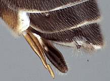
Anoncus. S1 very short, 0.25-0.35 times length of T1. T2 thyridium distinct in all but one of species examined; laterotergites of T2 and T3 completely separated by creases. Shallow transverse grooves present subapically on T1, T2, and sometimes T3 (Fig. 12); T1 and T2 coarsely sculptured. Ovipositor fairly short (Fig. 13), straight, with well-developed subapical, dorsal notch; ovipositor sheath (Fig. 13) not parallel-sided. Apex of female metasoma as in Fig. 13.

This description is modified from that of Townes, 1970: 127 and based on 3-4 species in the Texas A&M University Collection.

10133_mximage
1. Rhinotorus ha...
10121_mximage
2.Rhinotorus habitus
10170_mximage
3. Rhinotorus face and clypeus...
10119_mximage
4. Rhinotorus face and clypeus...
10132_mximage
5. Rhinotorus face and clypeus and mandibles...
10118_mximage
6.Rhinotorus mandibles
10130_mximage
7. Rhinotorus propodeum...
10136_mximage
8. Rhinotorus propodeum and metapleu...
10129_mximage
9.Rhinotorus wings
10131_mximage
10. Rhinotorus T1 dorsal...
10128_mximage
11.Rhinotorus T1 and S1
10135_mximage
12.Rhinotorus T1-T3
10134_mximage
13. Rhinotorus ovipositor and sheath...
 
Distribution
No referenced distribution records have been added to the database for this OTU.
Biology / Hosts
A large number of tenthredinid hosts have been reported for the species of Rhinotorus and the literature is summarized by Yu et al. (2012).
Map

There are no specimens currently determined for this OTU, or those specimens determined for this OTU are not yet mappable.

Acknowledgements
This page was assembled by Bob Wharton as part of a larger collaborative effort on the genera of Ctenopelmatinae. Page last updated May, 2015.

This work would not have been possible without the groundwork provided by Ian Gauld’s study of the Australian and Costa Rican faunas, and we are particularly grateful for his assistance in many aspects of this study. We also thank David Wahl of the American Entomological Institute, Andy Bennett of the Canadian National Collection, and Gavin Broad of The Natural History Museum, London for extended loans of the material used for this study and Dave Karlsson for access to material from the Swedish Malaise Trap Survey (trap 20, collection events 233 and 234). Matt Yoder provided considerable assistance with databasing issues, and our use of PURLs (http://purl.oclc.org) in this regard follows the example of their use in publications by Norm Johnson. Heather Cummins, Andrea Walker, Patricia Mullins, Caitlin Nessner, Mika Cameron, Karl Roeder, Amanda Ladigo, and Cheryl Hyde graciously assisted with image processing, formatting, and literature retrieval. This study was supported by the National Science Foundation’s PEET program under Grant No. DEB 0328922 and associated REU supplement #s DEB 0723663, 0923134, and 1026618.

This material is based upon work at Texas A&M University supported by the National Science Foundation under Grant Number DEB 0328922 with REU supplements DEB 0723663, 0923134, and 1026618. Any opinions, findings, and conclusions or recommendations expressed in this material are those of the author(s) and do not necessarily reflect the views of the National Science Foundation.