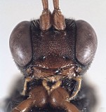
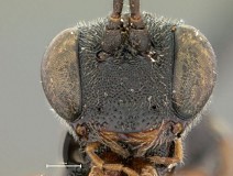
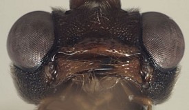
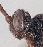
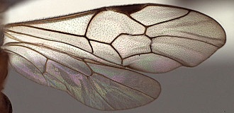
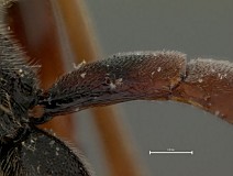
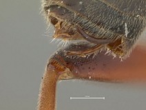
Clypeus with surface usually polished, sparsely punctate, and sparsely setose (Figs 4, 5); ventral margin varying from weakly to distinctly convex, the margin blunt (Fig. 4); epistomal sulcus impressed throughout, weak medially in some species (Fig. 5), very strongly impressed in others; clypeus in profile weakly protruding (Fig. 7). Face densely granular-punctate, frons and vertex often similarly sculptured, though vertex slightly less so in some species (Fig. 8); inner eye margins straight to weakly converging (Figs 4, 5). Malar space absent or nearly so (Figs 4, 5). Mandible (Fig. 6) strongly curved (Fig. 9), narrowing gradually from base to apex; dorsal tooth varying among species from equal to distinctly longer than ventral tooth; basal, transverse impression absent; ventral margin distinctly carinate. Ocelli small, distance from lateral ocellus to eye usually at least twice diameter of lateral ocellus (Fig. 8). Maxillary palp shorter than height of head, in some species only half head height; female antenna slightly to distinctly shorter than body. Hypostomal carina meeting occipital carina at base of mandible (Fig. 9); occipital carina complete. Epomia absent or very indistinct. Epicnemial carina usually not reaching anterior margin of mesopleuron (Figs. 10, 11), rarely appearing to do so. Notaulus usually absent (Fig. 10) or nearly so, rarely impressed on anterior declivity, extending posteriorly to level of tegula as barely detectable impressions. Groove between propodeum and metapleuron u-shaped, readily visible in lateral view; broad, u-shaped groove mid-dorsally between propodeum and metanotum also readily visible in lateral view; pleural carina usually well-developed, complete, but sometimes obscured by heavy sculpture; median and lateral longitudinal carinae usually well developed and complete; posterior transverse carina complete, anterior transverse variable, rarely complete, with costula and/or median portion absent, areola thus sometimes present and sometimes confluent with median basal area; petiolar area always well -defined, without median longitudinal carina. Hind femur moderately stout (Fig. 2) to somewhat more slender (Fig. 3); hind trochantellus (= second trochanter) flattened ventrally, the flattened portion at least weakly carinate on its lateral margin and distinctly carinate apically; apical margin of mid tibia often expanded into a distinct tooth similar to that of fore leg, though more rounded and less distinct in some species; apical comb on posterior side of hind tibia absent; posterior hind tibial spur short (Fig. 3), about 7x longer than maximum width at base; tarsal claws in at least some species pectinate at extreme base (difficult to see), pectination apparently absent in other species. Fore wing areolet absent in the five species examined, though
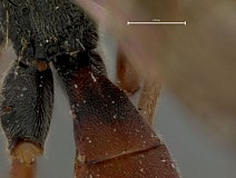
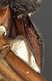
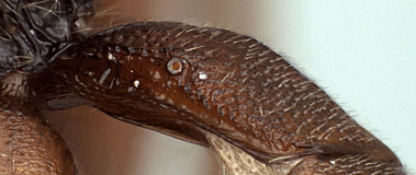
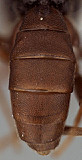
Townes (1970) indicated that it is sometimes present; stigma relatively narrow (Fig. 14), Rs+2r either arising near basal 0.3-0.4 of stigma or near midpoint. Hind wing (Fig. 14) with first abscissa of CU1 distinctly longer than 1cu-a. T1 (Figs 3, 15-18) short, broad, gradually expanded from base to apex, never parallel-sided (Fig. 15); weakly decurved to essentially straight in profile; dorsal carinae distinct but variable in length among species: extending nearly full length in some species, but only covering basal 0.5 in others; basal depression at dorsal tendon attachment broad, obvious; dorsal-lateral carina complete along full length of T1 in some species, partially obscured by heavy sculpture, especially posteriorly, in others; glymma present as a basal impression: broad in some species (Fig. 17), narrower in others (Fig. 16). S1 short, 0.35-0.4 times length of T1, not extending to level of spiracle. T2 thyridium absent (Fig. 19), T2 usually sculptured, minimally densely punctate; laterotergites of T2 and T3 separated by creases from median tergite. Ovipositor and sheath (Figs 20-21) narrow, upcurved; ovipositor relatively shorter than in
Pion, abruptly becoming needle-like apically from distinctly swollen base (Fig. 21); without dorsal, subapical notch.
The above description is modified from Townes (1970) and based on material in the Texas A&M University Collection, including at least one undescribed species from Texas.
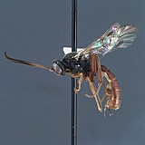
1.
Trematopygus habitus...
Trematopygus habitus
↰
↴
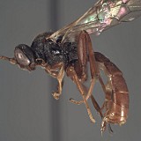
2.
Trematopygus habitus...
Trematopygus habitus
↰
↴
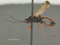
3.
Trematopygus apertor habitus...
Trematopygus apertor habitus
↰
↴
4.
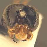
Trematopygus face...
Trematopygus face
↰
↴
5.Trematopygus apertor face
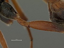
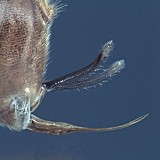
7.
Trematopygus head, la...
Trematopygus head, lateral view
↰
↴
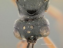
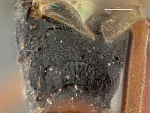
8.
Trematopygus apertor top of head...
Trematopygus apertor top of head
↰
↴
9.
Trematopygus back of hea...
Trematopygus back of head
↰
↴
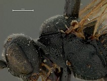
10.
Trematopygus apertor lateral...
Trematopygus apertor lateral
↰
↴
11.
Trematopygus mesop...
Trematopygus mesopleuron
↰
↴
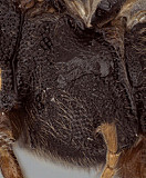
12.
Trematopygus apertor propodeum...
Trematopygus apertor propodeum
↰
↴
13.
Trematopygus apertor trochantellu...
Trematopygus apertor trochantellus
↰
↴
15.
Trematopygus apertor T1 dorsal ob...
Trematopygus apertor T1 dorsal oblique
↰
↴
16.
Trematopygus apertor T1 lateral...
Trematopygus apertor T1 lateral
↰
↴
17.
Trematopygus ...
Trematopygus T1
↰
↴
19.
Trematopy...
Trematopygus metasoma dorsal
↰
↴
20.
Trematopygus apertor ovipositor a...
Trematopygus apertor ovipositor and sheath
↰
↴
21.
Trematopygus ovipositor ...
Trematopygus ovipositor and sheath
↰
↴