Westwoodia gauldi Wharton and Roeder, 2008
Taxonomic History / Nomenclature
Westwoodia gauldi Wharton and Roeder, 2008: 9-11
Taxonomic Links
Westwoodia
Westwoodiini
Ctenopelmatinae
Westwoodiini
Ctenopelmatinae
Remarks
This species is known from only three specimens, which show sexual dimorphism in color pattern of the mesosoma (black in female, mostly orange in male) and the more basal origin of m-cu on the fore wing areolet in the male (Figs 17 and 18 above).
Diagnosis and Relationships

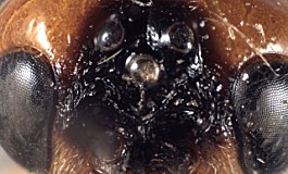
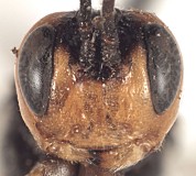
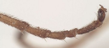
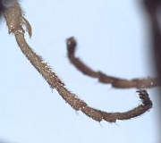
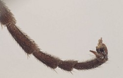
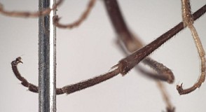
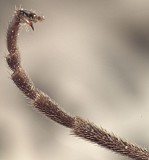
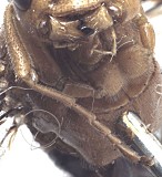
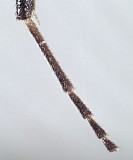
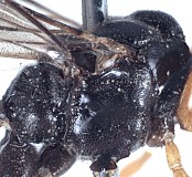
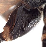
First flagellomere of antenna densely setose throughout (Fig. 1); interantennal flange (Fig. 2) tall, narrow, somewhat distant from ocelli, somewhat rectangular in profile; lateral swelling of frons with carinate inner margin (Fig. 3, arrow), especially near ocelli; face densely punctate medially (Figs 5-7), less densely punctate laterally; occipital carina complete, distinctly developed throughout (Fig. 8); female fore tarsus long, slender, and flattened (Figs 9, 10), hind tarsus (especially basitarsus) longer and more slender (Figs 11-14); Male tarsus as in Figs 15 and 16; fore wing (Figs 17, 18) with stigma dark brown and areolet present; 2m-cu usually arising near middle; fore tarsus yellow, hind tarsus brown to dark brown, mesosoma black and orange (male, Fig. 19) or black (female, Figs 20-22), metasoma dorsally, except for apical margin of terga, black.
Distinguished from all other species of Westwoodia by the combination of a densely punctate face, tall interantennal flange, strongly elevated median mesoscutal lobe, and sharply margined frontal depression. This species is most similar to some mainland populations of W. ruficeps in which the interantennal flange is not hemispherical in outline. In all populations of W. ruficeps, however, the tarsi, especially on the fore leg of the female, are very short and broad.
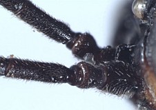
1.
Base of antenna showing pattern of s...
↴
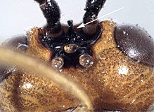
2.
Posterior view of head showing tal...
↴
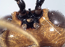
3.
Posterior view of head showing sha...
↴
4.
dorsal view of frons and ocelli showing car...
↴
5.Frontal view of head.
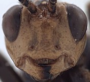
6.
Frontal view, top of head t...
↴
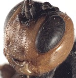
7.
Oblique view of head sh...
↴

8.
Back of head showing sharp...
↴
9.
Female fore tarsi, anterior vie...
↴
10.
Female fore leg, posterior ...
↴
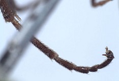
11.Female hind tarsi, anterior view.
12.Female hind tarsi, anterior view.
13.
Female hind tarsi, posterior vi...
↴
14.
Female hind tarsi, po...
↴
15.
Male fore leg, anteri...
↴
16.
Male hind leg, post...
↴
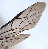
17.
Fore and hind wing, mal...
↴
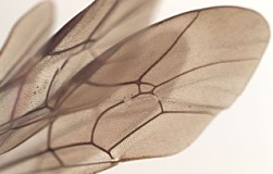
18.Fore wing, female.
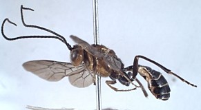
19.Habitus male.
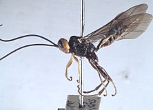
20.Habitus female.
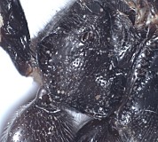
21.
Propodeum showing punctate ...
↴
22.
Mesosoma of female showing...
↴
23.
Hind coxa showing setal...
↴
Description
See monograph on Westwoodia by Wharton et al. (2008).
Distribution
Australia, New South Wales, ACT, and Victoria.
Distribution
No referenced distribution records have been added to the database for this OTU.
Biology / Hosts
Unknown
Biology and Behavior
Unknown
Map
There are no specimens currently determined for this OTU, or those specimens determined for this OTU are not yet mappable.
Label data
Holotype ♀(ANIC)
label = hand-written, black ink on white paper, 3 lines as follows:
BLACK MT
6 XII 29
J. W. EVANS.
label = hand-written, black ink on white paper, 3 lines as follows:
BLACK MT
6 XII 29
J. W. EVANS.
Paratypes
1♂, NSW, Tubrabucca, 10-23.i.1948, R.T.M.P. & A.N.B. (Victoria Museum); 1♂, VICTORIA, Launchins Place, 13.i.1913 (?), F.F.S. (Victoria Museum).
Actual paratype labels:
1) single label = printed, black ink on white paper, 3 lines as follows:
Tubrabucca, N.S.W.
Jan. 10-23, 1948
R.T.M.P. & A.N.B.
2) 2 labels
top label = hand-written, black ink on white paper, 3 lines as follows:
Launchins
Place. Vict
F.F.S. 13.1.13 (date hard to read, this is my best interpretation)
2nd label = printed, black ink on white paper, 2 lines as follows:
Ex. Coll.
Nat. Mus.
Acknowledgements
This page was assembled by Bob Wharton, and is part of a revision of the genus Westwoodia by Wharton, Karl Roeder, and Matt Yoder (Wharton et al. 2008). Kira Zhaurova analyzed the relationships among the Westwoodiini and Scolobatini as part of her M. S. thesis at Texas A&M University, completed in 2006. The material she borrowed for her thesis forms the basis for this revision of Westwoodia. We are grateful to Ken Walker (Victoria Museum, Melbourne) and John LaSalle (Australian National Insect Collection, Canberra) for extended loan of the specimens listed above. This material is based upon work conducted at Texas A&M University and supported by the National Science Foundation’s PEET program under Grant No. 0328922 and associated REU supplement # 0616851. Page last updated February, 2011.
This material is based upon work supported by the National Science Foundation under Grant Number DEB 0328922 with REU supplement 0616851.
Any opinions, findings, and conclusions or recommendations expressed in this material are those of the author(s) and do not necessarily reflect the views of the National Science Foundation.
