Type species: Gauldia levis Zhaurova, 2009.
Gauldia Zhaurova, 2009
Taxonomic History / Nomenclature
Gauldia Zhaurova, 2009 In: Zhaurova and Wharton (2009):
Remarks
The type species has a dark body with orange antennae and legs, largely hyaline wings, and white transverse apical bands on the metasomal tergites. It is further characterized by the pattern of setae and dense punctation on the face and clypeus, the tall inter-antennal flange.
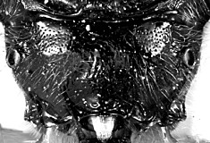
Species description: Female. Face deeply and densely punctate, slightly rugose medially and basolaterally. Prominent interantennal ridge extends posteriorly, splitting into 1 median and 2 lateral carinae beyond posterior margin of torulus. Ocelli fairly small and not distinctly raised, distance between eye margin and lateral ocellus 1.2 times distance between lateral ocelli. Frons polished medially amid carinae; vertex finely punctate throughout; distinct patch of matt sculpture extending diagonally from lateral ocellus to eye, with a distinct, weakly curved sulcus extending from lateral ocellus to eye. Pronotum uniformly punctate laterally. Mesopleuron quite densely punctate, lightly setose, with a fairly small impunctate area posteromedially. Metapleuron uniformly densely but finely punctate, setose. Propodeum with transverse carinae absent or indistinct.
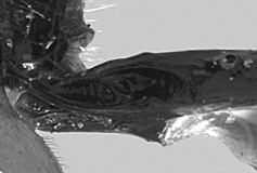
Gauldia shares two distinctive features of the petiole with Hypopheltes, namely the glymma with the deep part of the pit posterior to the dorsal tendonal attachment (dta) and the posteriorly excavated pit mid-dorsally behind the dta. The mid-dorsal pit and its posterior concavity limit the depth of the glymma on either side of the petiole, and the two structures are thus unlikely to be independent. Nevertheless, this is a complex character unique to these genera, strongly suggesting a close relationship. Gauldia is treated here as a separate genus (relative to Hypopheltes) largely on the basis of sculptural differences on the head and propodeum, the latter largely responsible for its intermediate placement between Hypopheltes and Westwoodia in our analyses (Zhaurova and Wharton 2009): 69-76. The genus is known from a single species; males are thus far unknown; Gauld (1984) treated this as an undescribed species under Hypopheltes.
Diagnosis and Relationships

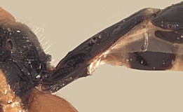
Gauldia shares with Hypopheltes characteristics of the petiole that distinguish them from all other westwoodiines. These include the deep, wide glymma (Figs 1, 2) and the tunnel-like posterior excavation of the pit behind the attachment of the dorsal tendon. Unlike the known species of Hypopheltes, there is a prominent interantennal flange in Gauldia, and the propodeum in Gauldia lacks the extensive carination found in Hypopheltes. The pale-colored, undoubtedly nocturnal species of Hypopheltes also have greatly enlarged ocelli compared to those of Gauldia.
Description
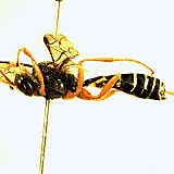
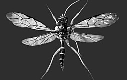
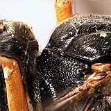
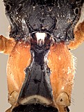
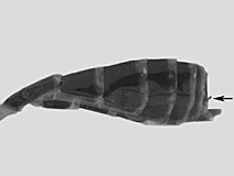
Length: body 12.7-13.3 mm (Figs 2, 3), fore wing 12-13 mm. Head: Clypeal margin blunt, very slightly thickened and weakly protruding medially, widely truncate (Fig. 1). Clypeus 3.4-3.5 times as broad as long, moderately protruding and weakly convex in profile, quite densely punctate, without a median transverse sulcus. Anterior tentorial pits small but distinct, epistomal sulcus distinct throughout, though less deeply impressed than in Hypopheltes. Malar space 0.5 times basal width of mandible, mandible with ventral tooth slightly longer than dorsal, sides parallel over apical 0.7. Face (Fig. 1) about twice broader than long, terminating dorsally with a small median tooth, face deeply, densely punctate. Interantennal area with a prominently elevated ridge. Frons concave immediately behind scape; densely setose, densely punctate and weakly convex adjacent eye, where it is slightly elevated above the eye. Widest diameter of torulus about 1.3 times widest diameter of median ocellus. Area between lateral ocelli depressed medially. Area behind ocelli regularly rounded. Antennae longer than body, with 41-42 flagellomeres. Length of first flagellomere about 0.8 times widest transverse diameter of eye, second flagellomere about as long as first. Occipital carina complete, joining hypostomal carina above base of mandible. Occiput much shorter mid-dorsally, only half the distance between lateral ocellus and occipital carina. Pronotum dorsally in profile distinctly concave, quite strongly depressed medially, expanded anteriorly, with truncate anterior margin, with indistinct transverse sulcus giving rise to wide, indistinct lateral groove of the pronotum (Fig. 4); without epomia or weak transverse ridge. Mesoscutum convex in profile, densely punctate; notauli (Fig. 4) deep, reaching posterior margin of mesoscutum or nearly so. Mesopleuron with broad, fairly sharply defined longitudinal depression for reception of femur. Metapleuron punctate. Propodeum moderately convex, and of moderate length (Fig. 9), distance between spiracles about 1.6 times mid-dorsal length, median longitudinal carina weak (Fig. 7), extending over about apical 0.4 of propodeum, pleural carina (Figs 6, 9) complete, distinctly elevated throughout, touching lateral rim of spiracle. Tibia and tarsus slender, longest hind tibial spur equal to greatest apical width of tibia, claws simple. Fore wing (Fig. 5) with areolet present, sessile; 3rs-m always slightly longer than 2rs-m; 1cu-a opposite or slightly distad base of Rs+M, almost vertical; marginal cell relatively slender; first subdiscal cell weakly explanate distally. Hind wing (Fig. 5) with first abscissa of Cu1 equal to 1cu-a, or nearly so; distal abscissa pigmented to wing margin, tubular for most of its length. Glymma deep (Figs 9, 10), originating as a narrow depression anterior to dorsal tendon attachment, expanding as a wide, discrete excavation posteriorad dorsal tendon attachment; T1 with spiracles protruding, S1 reaching 0.6-0.7 of distance to spiracle. Dorsal tendon attaches to the base of a deep pit; pit excavated in tunnel-like fashion posteriorly.
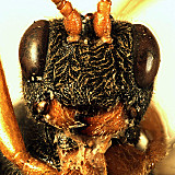
1.Gauldia levis face.
2.
Gauldia levis lateral ha...
↴
3.Gauldia levis dorsal habitus.
4.
Gauldia levis pronotum l...
↴
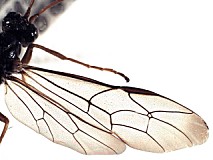
5.Gauldia levis wings.
6.Gauldia levis propodeum.
7.
Gauldia levis pr...
↴
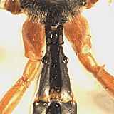
8.
Gauldia levis petiole, d...
↴
9.
Gauldia levis petiole, lateral view showin...
↴
10.Gauldia levis glymma.
11.
Gauldia levis female metasoma, ar...
↴
Distribution
Known only from a single locality in Victoria, Australia (holotype and two paratypes of the type species, Gauldia levis).
Holotype of G. levis: ♀, with four labels in addition to holotype label.
First label with three lines; first line: Bred from larvae second line: 1-vii-1959 third line: No. 525. Second label with four lines; first line: Australia second line: Victoria third line: Nerrina fourth line: M.F. Leask. Third label with four lines; first line: Adults = second line: Pseudoperga third line: belinda Kirby fourth line: ♀♂ recog Leask. Fourth label with two lines; first line: Brit. Mus. second line: 1959-440 (BMNH).
Distribution
No referenced distribution records have been added to the database for this OTU.
Biology / Hosts
The putative host, Pseudoperga belinda (Kirby) (Hymenoptera: Pergidae), is known only from an undetermined species of Eucalyptus, and has been collected in New South Wales, Tasmania, and South Australia in addition to Victoria (Schmidt and Smith 2006).
Map
There are no specimens currently determined for this OTU, or those specimens determined for this OTU are not yet mappable.
Acknowledgements
This page was assembled by Bob Wharton. It is part of a revision of the genera of Westwoodiini and Scolobatini conducted by Kira Zhaurova as part of her M. S. thesis in Entomology at Texas A&M University, completed in 2005, and subsequently published in part by Zhaurova and Wharton (2009).
This study would not have been possible without the groundwork provided by Ian Gauld’s study of the Australian fauna, and we are particularly grateful for his assistance in many aspects of this study. We also thank the following curators and researchers for extended loans of the material used for the revision of Scolobatini and Westwoodiini: David Wahl (AEIC), John LaSalle (ANIC), Ian Gauld and Gavin Broad (BMNH), Andy Bennett (CNC), Gabriel Melo (DZUP), Anders Albrecht and Pekka Malinen (FMNH), Ronald Zúñiga (INBio), Ken Walker (MVMA), Hege Vårdal (NHRS), Chris Burwell (QM), and Dave Furth (USNM). Matt Yoder provided considerable assistance along the way, particularly with databasing. Images used here were obtained through the combined efforts of Kira Zhaurova, Heather Cummins, and Patricia Mullins. Our use of PURLs (http://purl.oclc.org) follows the example of their use in publications by Norm Johnson. This material is based upon work conducted at Texas A&M University and supported by the National Science Foundation’s PEET program under Grant No. DEB 0328922 and associated REU supplement nos DEB 0723663 and DEB 0616851. Page last updated April, 2011.
This material is based upon work supported by the National Science Foundation under Grant Number DEB 0328922 with REU supplements DEB 0723663 and 0616851.
Any opinions, findings, and conclusions or recommendations expressed in this material are those of the author(s) and do not necessarily reflect the views of the National Science Foundation.
