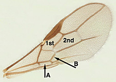
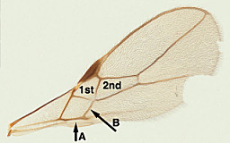
Couplet 4.
The Wharton Lab
The keys, text, and images presented here were initially developed as part of an NSF-sponsored PEET project, DEB0328922. This page is a joint project of NSF PEET grants NSF DEB-0328922; ideas and opinions expressed here are those of the authors, and not the NSF; best viewed with Firefox, Mozilla, or OS X Safari. This material is copyright Bob Wharton (2007 and onwards) and may not be reproduced without permission. 