Nehomaspis Heinrich, 1949: 15. Type species: Nehomaspis alpinus Heinrich by subsequent designation of Townes et al. (1965: 242). Synonymized by Townes et al. (1965: 242).
Homaspis Förster, 1869
Taxonomic History / Nomenclature
Homaspis Foerster, 1869: 198. Type species: Mesoleptus rufinus Gravenhorst by subsequent designation of Viereck (1914: 71)
Remarks
The Palaearctic species were revised by Kasparyan (2004); the Nearctic species by Barron (1990).
Twenty-one valid species are listed by Yu et al. (2012).
Twenty-one valid species are listed by Yu et al. (2012).
Diagnosis and Relationships

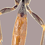
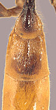
Homaspis is readily separated from Ctenopelma by the absence of a basal glymma. It is most readily separated from Xenoschesis by the presence of the lateral longitudinal carina (lacking in Xenoschesis). This thing doesn’t look anything at all like Ctenopelma even though they are supposedly in the same tribe. The Eastern Palaearctic Satous has the ventral tooth of the mandible about twice the length of the upper tooth whereas the two teeth are nearly equal in Homaspis. Homaspis is similar to Notopygus in these respects and the two are very similar to one another. T2 is a little shorter in Notopygus (Fig. 1 vs. Fig. 2), though this is not always apparent in males. The fore wing areolet is usually present in Notopygus and absent in Homaspis, but male Notopygus may have the areolet absent. Notopygus and Homaspis are most readily distinguished by differences in the female subgenital plate and relative position and shape of the ovipositor sheath. The subgenital plate is larger and rounded apicall in Notopygus but shorter, flatter, and apically subtruncate in Homaspis. The sheaths are compressed and dorsally directed in Notopygus but more or less depressed and dorso-posteriorly directed in Homaspis. In the relatively few species examined, the clypeus is rounded along the visible ventral margin in Notopygus but strongly impressed with a sharp ventral margin visible in Homaspis.
Description
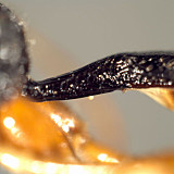
Frons without median horn or elevated carina. Clypeus short and wide, ventral 0.3-0.4 strongly impressed, with sharp, somewhat truncate to weakly concave ventral margin sometimes difficult to see, bluntly rounded and protruding at dorsal edge of impression; epistomal sulcus deep, distinctly separating clypeus from face. Malar space absent. Mandible moderately long, broad basally, not distinctly tapered apically, weakly curved; not or only weakly twisted; ventral tooth varying from equal to or very slightly longer than dorsal tooth. Lateral ocellus shorter than distance between ocellus and eye, though not strongly so. Maxillary palp shorter than head height; female antenna usually about as long as body, sometimes shorter; first flagellomere without tyloid. Hypostomal carina joining occipital carina slightly but distinctly above base of mandible; occipital carina complete. Epomia distinct to indistinct. Dorsal end of epicnemial carina distinctly separated from anterior margin of mesopleuron. Notaulus present anteriorly either as a short, broad, very shallow depression or a sharp, distinct groove fading distally to level of tegula. Propodeum and metanotum separated by broad, u-shaped groove or narrower, v-shaped groove middorsally in lateral view; large, distinct u-shaped notch visible between anterior end of lateral longitudinal carina of propodeum and adjacent part of metanotum in lateral view; pleural carina complete, well-developed; median and lateral longitudinal carinae of propodeum complete and well-developed throughout, areola not distinctly separated from basal area, the anterior transverse carina usually absent medially and always laterally (costula absent), petiolar area without median longitudinal carina, posterior transverse carina nearly always present and complete, rarely absent medially. Apical margin of mid tibia either not expanded or only slightly expanded as an angular tooth, always much less developed than that on fore tibia; comb on hind tibia absent to indistinct; hind tibial spurs of moderate length (longer than in Notopygus), straight to weakly curved distally; membranous flap on apical margin of fore tibia well-developed; all tarsal claws largely simple, pectinate only at extreme base and this usually difficult to see even though the species are large. Fore wing areolet nearly always absent (not present in any of the material examined for this redescription). Hind wing with first abscissa of CU1 usually shorter than 1cu-a, more rarely equal in length to 1 cu-a. T1 (Figs 1, 2) long and slender basally, gradually to abruptly widening posteriorly, apex < 4.0 (female) times wider than near base; dorsal carina sharp, distinctly elevated over basal 0.6-0.8, not extending to apex in material at hand; dorsal tendon attachment ending in small depression or not; dorsal-lateral carina sharp and distinct, extending from spiracle to apex; glymma absent but small, distinct pit and/or groove present just anterior to spiracle. S1 long, extending beyond middle to level of spiracle. T2 lateral carina sharp, strongly elevated; T2 thyridium absent; T2 size as in Fig. 2; laterotergites of T2 and T3 completely separated by creases. Ovipositor and sheath short, directed dorso-posteriorly to posteriorly; sheath somewhat flattened, depressed, apical portion of ovipositor distinctly narrow, without evident subapical notch in two species examined; female subgenital plate moderately large but not projecting, somewhat truncate posteriorly, often enclosed by lateral portions of tergum. Anal opening and cerci incompletely encircled by eighth metasomal tergum.
The above description is modified from Townes (1970) and was based largely on one North American and two eastern Palaearctic species in the Texas A&M University Collection.
Distribution
Holarctic.
Distribution
No referenced distribution records have been added to the database for this OTU.
Biology / Hosts
Detailed biologies for two species of Homaspis are given by Eichhorn (1988).
The following hosts and references for those hosts records are from Yu et al. (2005).
The following hosts and references for those hosts records are from Yu et al. (2005).
Acantholyda erythrocephala (Aubert 2000)
Acantholyda erythrocephala Pinus resinosa (Asaro and Allen 1999)
Acantholyda erythrocephala Pinus strobus (Asaro and Allen 1999)
Acantholyda luteomaculata (Barron 1990)
Acantholyda maculiventris (Barron 1990)
Acantholyda posticalis (Aubert 2000)
Cephalcia (Martinek 1989; Barron 1990)
Cephalcia abietis (Sedivy 1967; Jahn 1978; Eichhorn 1988; Martinek 1989, 1990; Kanecka 1993, 1995; Eichhorn and Bogenschutz 2000; Kasparyan 2002)
Cephalcia arvensis (Martinek 1991)
Cephalcia lariciphila (Aubert 2000)
Pamphilius depressus (Hinz 1961)
Pamphilius hypotrophicus (Strand 1915; Scheidter 1916; Hedwig 1962)
Pamphilius latifrons (Shaw et al. 2003)
Pamphilius ochrocera (Barron 1990)
Pamphilius vafer (Villemant 1982; Aubert 2000 )
Map
There are no specimens currently determined for this OTU, or those specimens determined for this OTU are not yet mappable.
Acknowledgements
This page was assembled by Bob Wharton as part of a larger collaborative effort on the genera of Ctenopelmatinae, with work on the Ctenopelmatini assisted by Matt Clark. Page last updated April, 2015.
This work would not have been possible without the groundwork provided by Ian Gauld’s study of the Australian and Costa Rican faunas, and we are particularly grateful for his assistance in many aspects of this study. We also thank the following curators for extended loans of the material used for this study: David Wahl of the American Entomological Institute and Andy Bennett of the Canadian National Collection. We also thank David Wahl for useful feedback throughout our study. Matt Yoder provided considerable assistance with databasing issues, and our use of PURLs (http://purl.oclc.org) in this regard follows the example of their use in publications by Norm Johnson. Heather Cummins, Matt Clark, Patricia Mullins, Caitlin Nessner, Amy James, and Cheryl Hyde graciously assisted us with image processing, formatting, and literature retrieval. This study was supported by the National Science Foundation’s PEET program under Grant No. DEB 0328922 and associated REU supplement nos DEB 0723663, 0822676, and 0923134.
This material is based upon work at Texas A&M University supported by the National Science Foundation under Grant Number DEB 0328922 with REU supplements DEB 0723663, 0822676, and 0923134. Any opinions, findings, and conclusions or recommendations expressed in this material are those of the author(s) and do not necessarily reflect the views of the National Science Foundation.
