Campodorus Foerster, 1868

Taxonomic History / Nomenclature
Campodorus Foerster, 1869: 213. Type species: Mesoleius melanogaster Holmgren, 1857. Subsequent designation by Perkins (1962: 412).

Phagesorus Foerster, 1869: 212. Type species: Tryphon caligatus Gravenhorst, 1829. Subsequent designation by Townes et al. (1965: 261). Synonymized by Townes et al. (1965: 261).

Cuboscopesis Heinrich, 1952: 1080. Type species: Cuboscopesis epachthoides Heinrich, 1952. Original designation. Synonymized by Townes et al. (1965: 261).

Remarks
Campodorus is the largest genus of Ctenopelmatinae Kasparyan (2005), with well over 100 valid species.
Diagnosis and Relationships
Campodorus has historically been challenging to separate from Mesoleius and Perkins (1962: 412) considered it a synonym of Mesoleius. Townes (1970) used the angle of inclination of the first abscissa of CU1 of the hind wing as the first character in his key couplet separating Mesoleius from Campodorus but the second character in this couplet, the shape of the clypeus, seems more reliable and is the character emphasized by Kasparyan ( 2003, 2005). We have followed Kasparyan in this regard. In Campodorus, the apical margin is blunt medially (Fig. 1) whereas in Mesoleius it is sharp (Fig. 2). Essentially, in Campodorus, a median, subapical bulge extends ventrally to obscure the sharp margin medially. The separation is not perfect, and some species are still difficult to place, but this is the most reliable character we have found.

In addition to the close resemblance to Mesoleius, there are gradations between species of Campodorus and those in at least six other genera (Townes 1970).

9954_mximage
1.Campodorus
10632_mximage
2.Mesoleius
 
Description
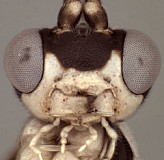
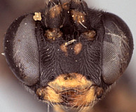
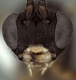
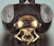
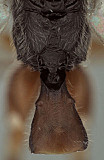
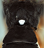
Clypeus (Figs 4-6) usually narrow, strongly bulging medially and subapically; ventral margin sharp except medially where sharp margin overlapped by blunt median bulge; ventral margin often bilobed (Fig. 5), with sharp lateral margins distinctly angled dorsally; epistomal sulcus varying from sharp and distinct to shallow and indistinct. Malar space in species examined equal to or shorter than half basal width of mandible (Figs 4, 5). Mandible (Figs 5, 6) usually relatively short and broad as in Figs 5 and 6, less commonly moderately long in material examined, curved, gradually narrowing from base to apex or more or less parallel-sided over apical half, ventral tooth varying from about equal in length to slightly shorter than dorsal tooth, with dorsal tooth usually broader. Inner eye margins parallel to weakly converging ventrally. Ocelli variable; commonly small, with maximum diameter of lateral ocellus distinctly shorter than distance between ocellus and eye. Female and male antennae at least as long as body in material examined, often longer than body; first flagellomere long and slender (Figs 1-3), nearly twice longer than second. Hypostomal carina joining occipital carina well above base of mandible; occipital carina complete. Epomia absent or sometimes present. Dorsal end of epicnemial carina nearly always extending to anterior margin of mesopleuron but sometimes weaker dorsally and distant from anterior margin in at least a few species; mesopleuron ventrally somewhat variable in sculpture from nearly smooth and polished to finely mat to more coarsely mat or rugulose, the punctures usually fine or obscured by mat sculpture, rarely more deeply punctate as in 
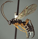
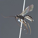
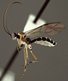
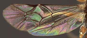
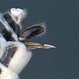
Otlophorus. Notaulus sharply impressed on anterior declivity and usually (80%) reaching anterior margin, usually distinct and sharply impressed on disk at least to level of tegula. Pleural carina usually well-developed, less commonly difficult to discern posteriorly; propodeal carinae about as in Fig. 7: lateral longitudinal carina of propodeum nearly always well-developed, complete or nearly so; median longitudinal carinae forming part of a broad (sometimes very large as in Fig. 8), rounded petiolar area then extending anteriorly as narrowly spaced, parallel-sided or converging ridges, rarely (5%) with median carinae absent anterior to petiolar area; petiolar area separated by transverse carina from narrower, triangular to parallel-sided areola, areola sometimes confluent with basal median area; otherwise, anterior and posterior transverse carinae absent. Legs usually with apical comb on posterior side of hind tibia poorly developed; hind tibial spurs usually long, slender (Fig. 3), less commonly shorter and stouter as in Fig. 1; all tarsal claws usually simple, less commonly pectinate. Fore wing (Figs 1, 9) with areolet absent. Hind wing with first abscissa of CU1 about as long as (Fig. 9) or more commonly much longer than (Fig. 1) 1cu-a. T1 (Figs 3, 7, 8) not long and slender; gradually widening posteriorly; basal depression for dorsal tendon attachment deep; dorsal carinae well-developed basally, often extending from base to or posteriorad level of spiracle; dorsal-lateral carina sharp and distinct from spiracle to apex; glymma deep, broad basally, narrowing posteriorly. S1 short to very short, 0.17-0.35 times length of T1. T2 thyridium present; laterotergites of T2 and T3 completely separated by creases. Ovipositor usually fairly short (Figs 2, 10), straight or weakly decurved, with deep, wide subapical, dorsal notch. Apex of female metasoma as in Fig. 10.

This description is modified from Townes (1970) and based on about 10 species in the Texas A&M University Collection.

9956_mximage
1. Campodorus habitus...
5188_mximage
2. Campodorus habitus...
9955_mximage
3. Campodorus habitus...
9952_mximage
4. Campodorus face and cl...
9954_mximage
5. Campodorus mandibles and...
9975_mximage
6.Campodorus mandibles
9953_mximage
7. Campodorus p...
9976_mximage
8. Campodorus propodeum ...
9911_mximage
9.Campodorus wings
5018_mximage
10. Campodorus ovipositor...
 
Distribution
No referenced distribution records have been added to the database for this OTU.
Map

There are no specimens currently determined for this OTU, or those specimens determined for this OTU are not yet mappable.

Acknowledgements
This page was assembled by Bob Wharton as part of a larger collaborative effort on the genera of Ctenopelmatinae. Page last updated May, 2015.

This work would not have been possible without the groundwork provided by Ian Gauld’s study of the Australian and Costa Rican faunas, and we are particularly grateful for his assistance in many aspects of this study. We also thank David Wahl of the American Entomological Institute and Andy Bennett of the Canadian National Collection for extended loans of the material used for this study and particularly Dave Karlsson for sending valuable material from the Swedish Malaise Trap Survey and Dmitry Kasparyan for discussions of generic limits. Matt Yoder provided considerable assistance with databasing issues, and our use of PURLs (http://purl.oclc.org) in this regard follows the example of their use in publications by Norm Johnson. Heather Cummins, Andrea Walker, Patricia Mullins, Caitlin Nessner, Amanda Ladigo, Mika Cameron, Karl Roeder, Danielle Restuccia, and Cheryl Hyde graciously assisted with image processing, formatting, and literature retrieval. This study was supported by the National Science Foundation’s PEET program under Grant No. DEB 0328922 and associated REU supplement #s DEB 0723663, 0923134, and 1026618.

This material is based upon work at Texas A&M University supported by the National Science Foundation under Grant Number DEB 0328922 with REU supplements DEB 0723663, 0923134, and 1026618. Any opinions, findings, and conclusions or recommendations expressed in this material are those of the author(s) and do not necessarily reflect the views of the National Science Foundation.