Westwoodia ruficeps Brullé, 1846

Taxonomic History / Nomenclature

Westwoodia ruficeps Brullé, 1846: 127-128.

Westwoodia ruficeps: Dalla Torre 1901: 310 (catalog); Roman 1912: 240-241, 292-293 (identity); Morley 1913: 101-102, 135-136 (redescription, figure, new distribution records); Viereck 1914: 152 (as type species of Westwoodia); Roman 1915: 3-5 (diagnosis, key, first description of male; new distribution records); Viereck 1921: 144 (type species listed); Townes et al. 1961: 213, 447 (catalog; key to genera); Townes 1970: 56-58 (redescription of genus; key to genera of Westwoodiini); Casolari and Casolari Moreno 1980: 59 (specimens in Spinola collection); Gauld 1984: 226-227, 233-234 (figures of species, redescription of genus, key to genera); Gupta 1987: 356 (catalog); Yu and Horstmann 1997: 456 (catalog).

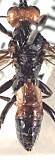
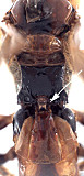
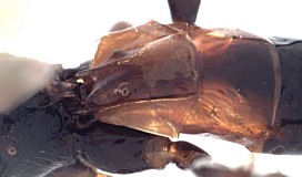
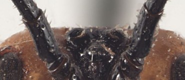
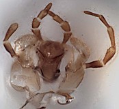
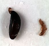
Westwoodia ruficeps was described from a single female specimen from Van Dieman’s Land (= Tasmania), “Collect. de M. Serville” (Brullé 1846). Many of the Brullé types from this work are in the Paris Museum, but it is also possible that Brullé returned the specimen to Audinet-Serville just before the Serville Collection was sold to Spinola in 1847. The Brullé Collection in the Paris Museum does not contain any specimens of Westwoodia, and Townes et al. (1961) stated that the type was lost. The Spinola Collection, however, contains two specimens of Westwoodia and although there is no locality label, they match in every respect material from the type locality. It is thus possible that one of these specimens is the holotype, but there is no way to be certain, especially since the Westwoodia specimens in the Spinola Collection apparently were obtained from Deyrolle rather than directly from Audinet-Serville (Casolari and Casolari Moreno 1980). Because populations from Tasmania are distinct in at least some respects from mainland populations, and since repeated attempts have failed to provide any evidence that the holotype still exists, we have designated a specimen from Hobart as neotype. The neotype was not selected from material in the Spinola collection because the two specimens examined are in poor condition.

Westwoodia ruficeps is the type species of the genus Westwoodia Brullé, 1846. Until recently (Gauld 1984), this was the only described species of Westwoodia.

Taxonomic Links
Westwoodia
Westwoodiini
Ctenopelmatinae
Remarks
VARIATION
We follow Gauld (1984) in interpreting Westwoodia ruficeps as a highly variable species. Some of the variation in morphology is readily categorized by population, but few populations are well represented in the material examined, with a result that the discussion here focuses on comparisons between material from Brisbane in southern Queensland and the type locality in Tasmania. A few comments are also offered on material from Victoria (primarily from Clunes) and Canberra (ACT). Unfortunately, no males from Tasmania were available for examination.

The Head: The transverse wrinkling on either side of the epistomal sulcus (shown in figures of the face in the Diagnosis and Relationships section) tends to be more extensive in material from Brisbane than Tasmania, and five specimens from Victoria and ACT lack this sculptural feature. In material from Victoria and ACT, the clypeus is often more uniformly truncate (rather than medially emarginate) and somewhat flatter (Fig. 1). The eye is variable in size, being longer and narrower in some individuals. The eye in lateral view is 1.0-1.15 x longer than the temple in specimens from Tasmania and the malar space in frontal view is 0.35-0.45 x the eye height. Comparable measures for Brisbane show non-overlapping ranges, with eye in lateral view 1.25-1.8 x longer than the temple and malar space 0.5-0.55 x eye height. Specimens from Tasmania also have more setae on the lower gena near the occipital carina whereas setae are virtually non-existent in this area in specimens from Brisbane. The median, elevated, blade-like flange on the frons tends to be longer and more evenly hemispherical in Brisbane material (Fig. 2) than material from Tasmania. In material from Tasmania, the flange more abruptly transitions to a low carina posteriorly as it extends to the median ocellus. Ten specimens from Victoria have a tall, more knob-like interantennal projection that does not extend posteriorly and is much more similar in structure to that of W. gauldi than to W. ruficeps. Setae on the first flagellomere were somewhat more decumbent and a little less sparse posteriorly than anteriorly in Tasmanian specimens relative to those from Brisbane, and proportions of the second and tenth (Fig. 4) flagellomeres were at the upper end of the range (2.75-3 Tasmania vs 2-2.8 Brisbane and 1.7-1.8 Tasmania vs. 1.2-1.7 Brisbane, respectively).

The Mesosoma: Specimens from Tasmania (Fig. 5) have the mesopleuron distinctly more setose than those from Brisbane (Fig. 6), especially in the depression below the subalar ridge. Material from intermediate areas is variable, with some more closely resembling Tasmania and most more closely approaching Brisbane. The pleural carina of the propodeum (Fig. 7) is generally better developed in material from Brisbane than from Tasmania. In the reared series from Brisbane (Fig. 8) and ACT, 2m-cu generally arises from the extreme apex of the areolet (0.85-0.95 x distance from base in females, 0.8-0.95 in males), while in a series from Victoria, 2m-cu arises from the basal 0.6-0.65. Material from Tasmania (Fig. 9) bridges the gap, ranging from 0.65-0.8. In this same Victoria series, 2cu-a consistently forms a weak angle with CU1. The areolet is variably petiolate within populations, as best exemplified by the long, reared series from Brisbane, where the stalk is very short, barely present in some and at least as long as the areolet in others. All specimens examined, regardless of locality, have short, stout fore tarsi with extensive fleshy lobes ventrally (Figs 10, 11). The hind legs (Figs 12-14) are more variable, however, being a little longer and more slender in material from Victoria, with hind leg basitarsus 4.3 (females) and 7.8-8.6 (males) x longer than wide. Hind tarsi of males from Victoria thus fall within the range for W. rodmani, but are still shorter than legs of other known species. The setal pattern of the posterior face of the second tarsomere of the hind leg (Figs. 13, 14) is distinctly denser in specimens from Tasmania and some specimens from Victoria than in the reared material from Brisbane and ACT.

Color Patterns: Specimens from Tasmania are darker (Figs 15, 16) than all mainland material examined with the exception of a few specimens from Victoria. Specimens from Brisbane nearly always have white to yellow fore tarsi (Figs 10, 11), pale apical flagellomeres (Fig. 3), yellow fore wing stigma (Fig. 8), wings more weakly infumate basally, and pale T1 and T2 (Figs 17-19). In approximately half of the mainland material, the mesosoma is largely pale yellow to orange (Fig. 19), with propodeum and metapleuron mostly to entirely black, rarely (1%) entirely pale. Leg color varies within populations to some extent. Even within material from Brisbane, specimens reared from pergids on Eucalyptus have a higher percentage of the lightest colored individuals than specimens reared from pergids on Melaleuca. The neotype female from Hobart, Tasmania and the two specimens from the Spinola collection completely match the color pattern of W. ruficeps in Brullé’s original description, including the bicolored flange protruding upwards between the antennae and posteriorly onto the frons.

In summary, three distinct populations are evident in the material at hand: one from Tasmania, one from Brisbane, and one from south-central Victoria. In specimens from the type locality (Tasmania), the body is consistently darker than in specimens from southern Queensland, the interantennal flange is less evenly hemispherical, and the mesopleuron is not as sparsely setose. Specimens from areas in between are variable in these features, with some intermediate states, but a clear north-south cline was not evident. The reared series from Canberra, for example, is generally more similar to material from Brisbane than material from Tasmania. Most specimens from Canberra have the color and setal pattern of material from Brisbane, and most are reared from the same host, but the shape of the interantennal flange is variable and often more like material from Tasmania. One of the specimens from Canberra, however, matches the 10 specimens from Victoria (Clunes, Meredith, and Ballarat) that are differentiated from all other specimens by the shape of the interantennal flange and to a lesser extent by the shape and sculpture of the clypeus.

It is quite possible that the material from south-central Victoria represents a separate species, but because there are some intermediates, we are reluctant to treat material from south-central Victoria as a separate species without more extensive collections from southeastern Australia to determine patterns of host utilization and whether there is any additional evidence of intergradation in form and color in this critical region. If the material examined from Victoria represents a second species then the possibility cannot be ignored that reared material from Canberra and Brisbane represent yet a third species that is sympatric with and may be host specific to Pergagrapta polita. The true W. ruficeps may thus be confined to Tasmania, outside the presently known distribution of P. polita. Host records are needed for material from Tasmania.

This is a moderately large genus that is confined to Australia. Only one species has been described. Some specimens in London were reared from pergid sawflies on Eucalyptus. One of these was used for the generic illustration.

1671_mximage
1. Frontal view of head, spec...
1670_mximage
2. Interantennal flange, spec...
1451_mximage
3. Pale apical flagellomeres, specimen fr...
1453_mximage
4. Dark flagellomeres from m...
3749_mximage
5. Anterior-lateral ...
3755_mximage
6. Anterior-lateral view of...
1583_mximage
7. Propodeum and metapleuron, s...
1581_mximage
8. Wings, specimen from Bri...
1835_mximage
9.Wings, specimen from Tasmania.
1429_mximage
10. Female fore leg showing ...
4136_mximage
11. Male for leg showing denser patter...
1843_mximage
12. Female hind leg ...
1674_mximage
13. Female hind leg showing de...
1836_mximage
14. Female hind leg showing dense s...
1829_mximage
15. Habitus, specimen from Tasmania, sh...
1830_mximage
16. Dorsa...
3802_mximage
17. Dor...
1838_mximage
18. Dors...
4111_mximage
19. Dorsal v...
1672_mximage
20. Dorsal-lateral view of petiole ...
 
Diagnosis and Relationships
First flagellomere of antenna sparsely setose (Figs 1, 2); interantennal flange (Figs. 1-3) tall, broad, extending to or nearly to ocelli, hemispherical in profile (Fig. 3); lateral swelling of frons lacking carinate inner margin (Fig. 4); face finely and sparsely punctate (Figs 5-7); occipital carina complete, distinctly developed throughout (Figs 9, 10); female fore tarsus short, broad, and flattened (Figs 12-14); male with first three tarsomeres somewhat longer (Figs 15, 16); hind tarsomeres longer than fore tarsomeres in both sexes (Figs 17-24); fore wing stigma dark brown (Fig. 25) to yellow (Fig. 26); fore wing areolet present, with 2m-cu usually arising near middle of apical half (Fig. 25), but arising from distal corner in some populations (Fig. 26); specimens from type locality with fore and hind tarsi dark brown to black, mesosoma and metasoma dorsally (Figs 27, 28), except for apical margin of terga, black.

Distinguished from other species of Westwoodia by the distinctive interantennal flange and short, broad, strongly flattened fore tarsus. The fore and hind tarsi of females (Figs 12-14, 17-21) and fore tarsus of males (Figs 15, 16) are shorter and broader than in other described species, and have much larger eversible lobes, covering most of the ventral surface on tarsomeres 2-4 (the visibility of these lobes unfortunately varies with specimen preparation). Male hind tarsus (Figs 22-24) is also shorter than in other species, but not appreciably broader. Specimens from the type locality (Tasmania) are darker, with fore tarsus (Fig. 14), apical flagellomeres (Fig. 27), and metasoma (Figs 28-30) black, with the fore wing stigma dark brown (Fig. 25). Specimens from southern Queensland nearly always have white to yellow fore tarsus (Figs 12, 13, 15, 16), the apical flagellomeres pale (Figs 33-35), the metasoma pale basally (Figs 36-40), and the fore wing stigma yellow (Fig. 26). Specimens from areas in between vary in coloration and the shape of the interantennal flange, but a clear north-south cline was not evident, perhaps because because relatively few specimens are available for study. Though variable, the dorsal carina of T1 tends to be better developed than in other species, often extending to the level of the spiracle as a distinct ridge (Fig. 37). The antennae also tend to be shorter, with fewer flagellomeres in this species.

3686_mximage
1. Frontal view of head showing sp...
3708_mximage
2. Sparsely setose first flag...
1670_mximage
3. Interantennal flange showi...
3713_mximage
4. Posterior view of hea...
1831_mximage
5. Frontal view. Specimen...
1833_mximage
6. Oblique view of face....
1432_mximage
7. Oblique view of face...
1450_mximage
8. Head in profile. Specime...
3721_mximage
9. Occipital carina (arro...
3400_mximage
10. Back of head, looking ...
3745_mximage
11. First f...
4114_mximage
12. Female fore tars...
1428_mximage
13. Female fore tarsi, anter...
4116_mximage
14. Female fore tarsi. Specimen fro...
1433_mximage
15. Male fore tarsi, posterior view...
1434_mximage
16. Male fore tarsi, anterior view. Br...
1430_mximage
17. Female hind tarsi, poste...
1674_mximage
18. Female hind tarsi, posteri...
1836_mximage
19. Female hind tarsus, posterior v...
1431_mximage
20. Female hind tarsi, anter...
1843_mximage
21. Female hind tars...
1745_mximage
22. Male hind tarsi, posteri...
1436_mximage
23. Male hind tarsi, anter...
1744_mximage
24. Male hind tarsi...
1835_mximage
25. Right fore and hind wing showin...
4118_mximage
26. Fore and hind wings showin...
1842_mximage
27. Dorsal view showing anten...
1830_mximage
28. Dorsa...
4109_mximage
29. Dorsal view, ...
1829_mximage
30. Female habitus. Specimen from Tasma...
1675_mximage
31. Female habitus. Specimen f...
1676_mximage
32. Male habitus. Specimen from B...
1457_mximage
33. Antenna showing length relative to body. ...
1452_mximage
34. Subapical flagellomeres showing...
1451_mximage
35. Pale apical flagellomeres. Brisbane sp...
1838_mximage
36. Dors...
1582_mximage
37. Sculpture ...
1585_mximage
38. Dorsal ...
3802_mximage
39. Dor...
3803_mximage
40. Dors...
3685_mximage
41. Dorsal view showing short but deep notau...
1586_mximage
42. Apex of metasoma showi...
1584_mximage
43. Lateral view of thorax sh...
4155_mximage
44. Hind coxa showing unifo...
3402_mximage
45.Maxilla and labium.
3397_mximage
46. Egg of Westwoodia (right) ...
 
Description
See monograph on Westwoodia by Wharton et al. (2008).
Distribution
Australia
The species was originally described from a single specimen collected in Tasmania (Brullé 1846). Subsequent authors have interpreted the species widely, ranging from Tasmania north to northern Queensland and west into South Australia (Gauld 1984). Though this species occurs from Queensland to Tasmania in eastern Australia, we have seen only a few individuals from NSW, and relatively few localities are represented by the material at hand.

Roman (1915)

recorded a female from Fremantle (just south of Perth) and noted that since it was previously known only from Tasmania, this new record suggests that W. ruficeps is distributed across the whole of South Australia. We have not seen Roman’s specimen from Fremantle but two specimens in the Western Australian Museum were collected from the vicinity of Perth. Morley (1913) records specimens from Adelaide and near Melbourne as W. ruficeps. Unfortunately, his redescription is likely based on two different species (see comment under the W. longipes species page). Thus, distribution of W. ruficeps remains to be confirmed outside the southwestern and southwestern parts of Australia and all along the eastern margin of the continent.

Distribution
No referenced distribution records have been added to the database for this OTU.
Biology / Hosts
All known hosts belong to sawflies of the subfamily Perginae (Hymenoptera: Pergidae). We examined 31 specimens from Brisbane reared from Pergagrapta polita (Leach) (Pergidae) collected as larvae feeding on leaves of Eucalyptus propinqua H. Deane & Maiden and Melaleuca quinquenervia (Cav.) S. T. Blake. An additional 11 specimens from Canberra were also reared from P. polita but there is no host plant information on the labels. There is also one specimen from Clunes (Victoria) reared from Pergagrapta spinolae (Westwood), also without host plant data.

The following records require some confirmation because of uncertainties regarding the identity of the host or the host plant. Gauld (1984) lists a record for Pseudoperga sp., and the label on this specimen indicates Eucalyptus pauciflora as the plant host. Unfortunately there is no locality data on the label and six species of Pergidae (including two in Pseudoperga Guérin-Méneville and two in Pergagrapta Benson) are known from this host plant (Schmidt and Smith 2006). Since some species now in Pergagrapta were formerly included in Pseudoperga (Schmidt and Smith 2006), the host for this record cannot be further pinpointed. There is also a specimen from Clunes that was observed attacking “probably” Pergagrapta bicolor (Leach) on Eucalyptus dives Schauer. Eucalyptus dives is not a known host for P. bicolor, but two species of Pseudoperga are known from this host (Schmidt and Smith 2006).

Biology and Behavior
Virtually nothing is known of the biology and behavior of members of the genus Westwoodia, other than the host records listed here and under the species page for Westwoodia romani. Three short anecdotal accounts are available, two from labels and one from Lewis (1839). A female specimen from Ballarat, Victoria, was collected “in contact, a stride, clinging tenaceously to probably Pergagrapta bicolor. In situ on Eucalyptus dives, “blue peppermint” in bush.” A male specimen from ACT was collected “in copulo on Acacia melanoxylon overhanging creek.” Lewis (1839) records Ichneumonidae attacking pergid larvae, which “defend themselves, sometimes effectively, with exudates.”
Map

There are no specimens currently determined for this OTU, or those specimens determined for this OTU are not yet mappable.

Label data
Neotype ♀ (ANIC)
top label = printed, black ink on white paper, 3 lines as follows:
C.E. Cole
Hobart
4.11.16/2 [the /2 is hand-written]

2nd label = printed, black ink on white paper, 2 lines as follows:
Ex. Coll.
Nat. Mus.

3rd label = hand-written, black ink on white paper, 3 lines as follows:
WESTWOODIA
DET. IN MUS
COLL.

Additional material examined:
ACT 8♀, 3♂ (MVMA) Black Mt, 28.i-10.iii.1937, Bred from Perga[grapta] polita larvae; 1♂ (MVMA) Black Mt., 19.ii.1936, A.D. Butcher; 1♀ (CNCI) Black Mt., 15-21.ii.1999, G. Gibson; 1♂ (ANIC) Brindabella Ra, 3.iii.1984, J. A. Vranjic and D. Horan ex Acacia melanoxylon overhanging creek, in copulation; 3♀ (ANIC) Canberra, 15.iv.1958, 4.ii.1959, and16.ii.1960, E. F. Riek; 1♀ (TAMU), Canberra, 4-10.i.1999, R. Wharton.

NEW SOUTH WALES 1♂ (BMNH) Casula, 11.ii.1958, M. I. Nikitin; 1♀ (ANIC) Wilson’s Valley, 36.12S, 148.43E, 28.ii.1974, P. Morrow, predator on Pseudoperga sp. larvae.

QUEENSLAND 1♂ (BMNH); 11♀, 5♂ (QMBA) Brisbane, Deagon, 27º20’S 153º04’E, 8.×.1997, S. Schmidt, larvae on Melaleuca quinquenervia, ex Pergagrapta nr. polita; 12♀, 3♂ (QMBA) Brisbane, Deagon, 27º40’S 152º51’E, 21.viii and 30.ix.1997, S. Schmidt, larvae on Eucalyptus propinqua, ex Pergagrapta polita; 1♂ (MVMA) Cape York, French; 1♀(QMBA) Chelmer, 7.5 km SSW Brisbane, 18.viii.1992, M. Purcell; 1♀ (AEIC) Mt. Nebo, viii; 1♂ (AEIC) Nord-Queensland, E. Heyne; 1♂ (BMNH) Stanthorpe, iv.1927, H. Jarvis.

TASMANIA 3♀ (ANIC), Hobart, 15.×.1916. (Same series as neotype.)

VICTORIA 1♀ (USNM) F. M. Baker; 2♀ (ANIC) Ballarat, 28.v.1957, M.F. Leask; 1♂ (BMNH) Ballarat, emerged 11.vii.1957, M.F. Leask, foodplant of Symphyta, Eucalyptus; 1♀ (BMNH) Ballarat, Glen Park, 11.xi.1956, M.F. Leask, actually in contact, a stride, clinging tenaceously to probably Pergagrapta bicolor, on Eucalyptus dives, “blue peppermint”; 4♂ (BMNH) Clunes, emerged iv-vi.1957, 7, 14&18.iii.1958, M.F. Leask, ex pergid/Symphyta on Eucalyptus; 1♀ (BMNH) Clunes, 10.ii.1959, M.F. Leask; 1♀ (BMNH) Clunes, 25.v.1959, M.F. Leask, bred from larvae, adults Pergagrapta spinolae; 1♀ (BMNH), Lamplaugh reservoir, 24.iii.1958, bred from larvae, M.F. Leask; 1♀ (MVMA) Meredith, 12.ii.1959, A. N.; NO/UNCERTAIN LOCALITY 5♀, 5♂, 1? (ANIC, BMNH, MVMA); 1♀ (ANIC) Bulls HD, E. Pauci; 1♀ (ANIC) E. pauciflora (PR), 6.iii.1973, parasitic on Pseudoperga larva; 2♀ (MRSN).

WESTERN AUSTRALIA 1♂ (WAMP) Darlington, vii.1969, G.M. Lowe; 1♀ (WAMP) Glen Forrest [sic], 25.viii.1976, S.M. Postmus, feeding at flowers of Hakea amplexicaulis.

Additional references
Casolari, C. & Casolari Moreno, R. (1980) Cataloghi I – Collezione Imenotterologica di Massimiliano Spinola. Museo Regionale di Scienze Naturali, Torino, 165 pp.

Gupta, V. K. (1987) The Ichneumonidae of the Indo–Australian area (Hymenoptera). A synonymic catalogue of the taxa described through 1985 together with a bibliography, 1960–1985 (Part 1. Subfamilies Pimplinae to Mesochorinae). Memoirs of the American Entomological Institute, 41(1), 1–597.

Lewis, R. H. (1837) Case of maternal attendance on the larva by an insect of the tribe of Terebrantia, belonging to the genus Perga, observed at Hobarton, Tasmania. Transactions of the Entomological Society of London, 1, 232–234.

Lewis, R. H. (1839) Various observations on the natural history and entomology of Van Diemen’s Land. Proceedings of the Entomological Society of London, 2, xliv–xlv.

Morrow, P. A., Bellas, T. E. & Eisner, T. (1976) Eucalyptus oils in the defensive oral discharge of Australian sawfly larvae (Hymenoptera: Pergidae). Oecologia, 24, 193–206.

Roman, A. (1912) Die Ichneumonidentypen C. P. Thunbergs. Zoologiska Bidrag fran Uppsala, 1, 229–293.

Schmidt, S. & Smith, D. R. (2006) An annotated systematic world catalogue of the Pergidae (Hymenoptera). Contributions of the American Entomological Institute, 34(3), 1–207.

Weinstein, P. & Austin, A. D. (1995) Primary parasitism, development and adult biology in the wasp Taeniogonalos venatoria Riek (Hymenoptera: Trigonalyidae). Australian Journal of Zoology, 43, 541–555.

Yu, D. S. & Horstmann, K. (1997) A catalogue of World Ichneumonidae (Hymenoptera) Part 1: Subfamilies Acaenitinae to Ophioninae. Memoirs of the American Entomological Institute, 58(1), 1–763
.
Yu, D. S., van Achterberg, K. & Horstmann, K. (2005) World Ichneumonoidea 2004. Taxonomy, biology, morphology and distribution. Taxapad 2005.

Zhaurova, K. (2006) A revision of Physotarsus Townes, With a Preliminary Phylogenetic Analysis of Scolobatini (Hymenoptera: Ichneumonidae: Ctenopelmatinae). M.S. Thesis Texas A&M University, College Station, Texas, 162 pp.

Acknowledgements
This page was assembled by Bob Wharton, and is part of a revision of the genus Westwoodia by Wharton, Karl Roeder, and Matt Yoder (Wharton et al. 2008). Kira Zhaurova analyzed the relationships among the Westwoodiini and Scolobatini as part of her M. S. thesis at Texas A&M University, completed in 2006. The material she borrowed for her thesis forms the basis for this revision of Westwoodia. We are grateful to David Notton, Chris Burwell, Ken Walker and John LaSalle for the extended loan of the specimens listed above. This material is based upon work conducted at Texas A&M University and supported by the National Science Foundation’s PEET program under Grant No. 0328922 and associated REU supplement # 0616851.

We also thank John Goolsby and Chris Burwell for assistance in obtaining valuable reared material during our visit to Brisbane, to Stephan Schmidt, who reared the longest series available to us, and who also provided important information on hosts, and to Nicole Fisher and Aubrey Colvin for assistance with literature. We also thank Jacques Dubois for checking the Paris Museum for Brullé’s type, Andy Polaszek for comments on neotype designations, Guido Pagliano for information on the Spinola collection, and Jim Woolley for hand-carrying delicate material to and from BMNH. Page last updated April, 2011.

This material is based upon work supported by the National Science Foundation under Grant Number DEB 0328922 with REU supplement 0616851.
Any opinions, findings, and conclusions or recommendations expressed in this material are those of the author(s) and do not necessarily reflect the views of the National Science Foundation.