Megaceria Szépligeti, 1908
Taxonomic History / Nomenclature
Megaceria Szepligeti, 1908: 323. Type species: Megaceria opheltes Szepligeti, 1908. Monobasic.
Remarks
In his revision of the Australian Ichneumonidae, Gauld (1984: 236) identified two distinct species groups within Megaceria: the opheltes group contains M. opheltes Szépligeti, 1908 and M. pagana (Morley, 1913); the rufiventris group contains M. rufiventris (Brullé, 1846) and, according to Gauld (1984), at least 10 undescribed species. The two groups have remarkably different characteristics (as detailed in the description given on this webpage), and were they from the Holarctic Region, would probably have been described as separate genera.
There are only three valid species, known only from Australia:
Megaceria opheltes Szepligeti, 1908
Megaceria pagana (Morley, 1913)
Megaceria rufiventris (Brulle, 1846)
Diagnosis and Relationships

Confusion surrounding the identity and placement of Megaceria was clarified by Gauld (1984), who placed Megaceria in the Euryproctini based on the elongate petiole without glymma and lack of characteristics specific to westwoodiines and scolobatines such as a tyloid on the first flagellomere.
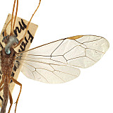
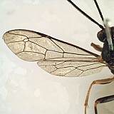
Denticeria and Megaceria were the only two Australian genera placed in Euryproctini by Gauld (1984). Gauld (1984) clearly differentiated these two from other Australian Ctenopelmatinae, but did not differentiate them from other, non-Australian Euryproctini. The distinctive distal broadening of the first subdiscal cell of the fore wing and the exceptionally short first abscissa of CU1 in the hind wing (as in Fig. 1) characterize these two genera relative to other euryproctine genera as treated in the key by Townes (1970). Denticeria has a shorter, broader T1 than does Megaceria, and the laterotergites are not separated by a crease on T3 in Megaceria.
Members of the rufiventris group closely resemble those westwoodiines without a distinct glymma, especially Pergaphaga (because of the reduced propodeal sculpture relative to Dicytopheltes, the other westwoodine without a distinct glymma). Megaceria is most easily separated from westwoodiines, including Pergaphaga, by the relatively longer first flagellomere lacking tyloids and by the hind wing venation.
1.Megaceria
Description
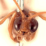
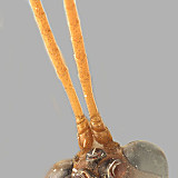
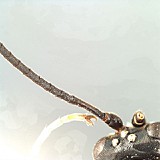
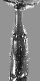
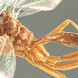
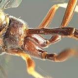
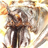
Clypeus tall and relatively narrow (Fig. 3); ventral margin almost evenly but shallowly convex in rufiventris group, more distinctly truncate to very shallowly and broadly concave medially and more sharply angled laterally in opheltes group (Fig. 3), margin blunt in both groups; epistomal sulcus present, usually not very distinct, broader and often more shallow in opheltes group; clypeus protruding in profile. Inner eye margins parallel or nearly so. Malar space varying from absent or nearly so in opheltes group (Fig. 3) to distinct, nearly half basal width of mandible in rufiventris group; malar sulcus absent. Mandible shorter, broader, with ventral and dorsal tooth of equal length in rufiventris group, mandible slightly longer and narrower, with ventral tooth slightly longer than dorsal tooth in opheltes group (Fig. 3). Ocelli variable: large, with lateral ocellus distinctly longer than distance between ocellus and eye in opheltes group (Fig. 4), but lateral ocellus much smaller, shorter than distance between ocellus and eye in rufiventris group (Fig. 5). Maxillary palps shorter than head height in rufiventris group, about equal to height of head in opheltes group; female antennae varying from approximately equal to body length in rufiventris group to longer than length of body in opheltes group; first flagellomere without tyloids, at least twice longer than second. Occipital carina complete dorsally and laterally, meeting hypostomal carina distinctly above base of mandible in rufiventris group, meeting or slightly effaced just before meeting hypostomal carina at base of mandible in opheltes group. Epicnemial carina distinctly removed from anterior margin of mesopleuron, terminating dorsally in rounded transverse ridge that sharply delimits a median longitudinal furrow extending across the middle of the mesopleuron. Notaulus weak in most specimens of opheltes group, though often extending nearly to posterior margin, very well developed in one species of rufiventris group as deep, narrow, unsculptured groove extending almost full length of mesoscutum, converging posteriorly on either side of elevated knob, weaker in other species examined. Lateral and posterior margins of scutellum completely to incompletely carinate in rufiventris group, completely acarinate in opheltes group. Without distinct, pionine-like, u-shaped groove between propodeum and metanotum in lateral view; pleural carina complete, usually well-developed; transverse and longitudinal propodeal carinae absent in at least one species of rufiventris group, posterior transverse carinae otherwise complete and well developed (Fig. 7), median longitudinal carinae usually present as narrowly spaced parallel ridges of varying length between posterior transverse carina and basal median depression, lateral longitudinal carinae absent anteriorly, ususally present and well developed posteriorad posterior transverse carina. Hind femur slender; apical margin of mid tibia not expanded into a distinct tooth similar to that of fore leg; apica; comb on inner side of apex of hind tibia absent, though this area covered with setae in opheltes group; tibial spurs not short and broad; hind leg with fifth tarsomere long and curved in rufiventris group, shorter, stouter in opheltes group; tarsal claws large, simple, strongly curved apically, with about 3 very long, widely spaced, slightly thickened setae along basal portion, these not darkened or sclerotized as in pectinate claws. Fore wing stigma narrow in opheltes group to very narrow in rufiventris group (Fig. 2), r arising slightly basal middle of stigma; fore wing areolet large, somewhat in the form of a lop-sided rectangle; first subdiscal cell usually enlarging proximally to distally (Figs 1, 2); 1m-cu&M straight in rufiventris group (Fig. 2), distinctly bent in opheltes group (Fig. 1); fore wing A straight or nearly so in rufiventris group (Fig. 2), distinctly bowed towards margin in opheltes group (Fig. 1). Hind wing with distal abscissa of CU1 slightly variable, arising near junction between CU1 and M or, in at least one species, arising from M slightly distad the junction. T1 long, slender, without glymma or dorsal carinae; no basal depression at dorsal tendon attachment, dorsal tendon attachment actually elevated above surface (Figs. 8, 9); dorsal-lateral carina absent between spiracle and apex of petiole; T1 and S1 fused, the suture absent; S1 long but variable, extending just posteriorad level of spiracle in most members of the opheltes group, extending well beyond spiracle in rufiventris group, in one species nearly equal distance between spiracle and dorsal tendon attachment. T2 thyridium absent. Ovipositor straight or nearly so, short, broad, with deep subapical notch; ovipositor sheath straight, relatively narrow, bluntly rounded apically, densely setose throughout in opheltes group bare or nearly so except at apex in rufiventris group; female subgenital plate densely setose in opheltes group, very sparsely setose in rufiventris group. Male parameres large, protruding, broadly truncate apically, never strongly narrowed; aedeagus variously spinose.
The above description is extensively modified from Gauld (1984) and is based on numerous specimens borrowed from museums indicated in the acknowledgements. The description of Megaceria by Townes (1970) actually refers to Pergaphaga Gauld, 1984. Townes (1970) based his description on a misidentified specimen that Gauld (1984) subsequently recognized as an undescribed genus. Short (1978) similarly misidentified Megaceria and thus his description of the larva pertains to Pergaphaga.
Distribution
No referenced distribution records have been added to the database for this OTU.
Biology / Hosts
The lectotype of Megaceria pagana Morely was reputed to have been reared from a lepidopterous pupa. Townes (1970) regarded the host record as suspect, since members of the subfamily are almost exclusively parasitoids of sawflies. However, Gauld (1984) subsequently confirmed Morley’s (1913) record by examining several additional specimens with lepidopteran host records.
Map
There are no specimens currently determined for this OTU, or those specimens determined for this OTU are not yet mappable.
Acknowledgements
This page was assembled by Bob Wharton as part of a larger collaborative effort on the genera of Ctenopelmatinae, with assistance by Kira Zhaurova on the Australian ctenopelmatines. Page last updated April, 2015.
This work would not have been possible without the groundwork provided by Ian Gauld’s study of the Australian and Costa Rican faunas, and we are particularly grateful for his assistance in many aspects of this study. We also thank the following curators and researchers for extended loans of the material used for this study: David Wahl of the American Entomological Institute, John LaSalle of the Australian National Insect Collection, Andy Bennett of the Canadian National Collection, Ian Gauld and Gavin Broad of The Natural History Museum, London, Ken Walker of the National Museum of Victoria (Australia), Chris Burwell of the Queensland Museum, Australia, and Bob Kula of the US National Museum of Natural History. We also thank David Wahl for useful feedback throughout our study. Matt Yoder provided considerable assistance with databasing issues, and our use of PURLs (http://purl.oclc.org) in this regard follows the example of their use in publications by Norm Johnson. Heather Cummins, Mika Cameron, Patricia Mullins, Caitlin Nessner, Danielle Restuccia, and Cheryl Hyde graciously assisted us with image processing, formatting, and literature retrieval. This study was supported by the National Science Foundation’s PEET program under Grant No. DEB 0328922 and associated REU supplement nos DEB 0522836, 0616851, 0723663, and 0923134.
This material is based upon work at Texas A&M University supported by the National Science Foundation under Grant Number DEB 0328922 with REU supplements DEB 0522836, 0616851, 0723663, and 0923134. Any opinions, findings, and conclusions or recommendations expressed in this material are those of the author(s) and do not necessarily reflect the views of the National Science Foundation.
